
“Three Methods and Three Points” regulates p38 mitogen-activated protein kinase in the dorsal horn of the spinal cord in a rat model of sciatic nerve injury
2017-01-21 03:33:16XinGuoTianyuanYuWongStevenWenduanJiaChiMaYanhongTaoChaoYangTaotaoLvShuaiWuMengqianLuJialiLiu
中國(guó)神經(jīng)再生研究(英文版) 2016年12期
Xin Guo, Tian-yuan Yu,, Wong Steven, Wen-duan Jia, Chi Ma, Yan-hong Tao, Chao Yang, Tao-tao Lv, Shuai Wu, Meng-qian Lu, Jia-li Liu
1 College of Acupuncture-Moxibustion and Tuina, Beijing University of Chinese Medicine, Beijing, China
2 College of Taditional Chinese Medicine, Beijing University of Chinese Medicine, Beijing, China
“Three Methods and Three Points” regulates p38 mitogen-activated protein kinase in the dorsal horn of the spinal cord in a rat model of sciatic nerve injury
Xin Guo1, Tian-yuan Yu1,*, Wong Steven1, Wen-duan Jia1, Chi Ma1, Yan-hong Tao1, Chao Yang1, Tao-tao Lv1, Shuai Wu1, Meng-qian Lu1, Jia-li Liu2
1 College of Acupuncture-Moxibustion and Tuina, Beijing University of Chinese Medicine, Beijing, China
2 College of Taditional Chinese Medicine, Beijing University of Chinese Medicine, Beijing, China
How to cite this article:Guo X, Yu TY, Steven W, Jia WD, Ma C, Tao YH, Yang C, Lv TT, Wu S, Lu MQ, Liu JL (2016) “Three Methods andThree Points” regulates p38 mitogen-activated protein kinase in the dorsal horn of the spinal cord in a rat model of sciatic nerve injury . Neural Regen Res 11(12):2018-2024.
Open access statement:This is an open access article distributed under the terms of the Creative Commons Attribution-NonCommercial-ShareAlike 3.0 License, which allows others to remix, tweak, and build upon the work non-commercially, as long as the author is credited and the new creations are licensed under the identical terms.
Funding:This study was supported by the National Natural Science Foundation of China, No. 81373759; the Natural Science Foundation of Beijing of China, No. 7142097.
Graphical Abstract
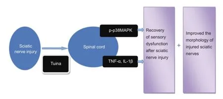
Pattern of tuina regulates the dorsal horn of the spinal cord of sciatic nerve injury
Tuina is a traditional Chinese treatment for sensory disturbances caused by peripheral nerve injury and related diseases. Our previous studies showed that tuina regulates relevant regions and indices of the spinal dorsal horn using the Dian, Bo, and Rou method in Yinmen (BL37), Yanglingquan (GB34), and Weizhong (BL40). Treatment prevents muscle atrophy, protects spinal cord neurons, and promotes sciatic nerve repair. The mechanisms of action of tuina for treating peripheral nerve injury remain poorly understood. This study established rat models of sciatic nerve injury using the crushing method. Rats received Chinese tuina in accordance with the principle of “Three Methods and Three Points,” once daily for 20 days. Tuina intervention reduced paw withdrawal latency and improved wet weight of the gastrocnemius muscle, as well as promoting morphological recovery of sciatic nerve fbers, Schwann cells, and axons. The protein expression levels of phospho-p38 mitogen-activated protein kinase, tumor necrosis factor-α, and interleukin-1β also decreased. These fndings indicate that“Three Methods and Three Points” promoted morphological recovery and improved behavior of rats with peripheral nerve injury.
nerve regeneration; tuina; Three Methods and Three Points; phospho-p38 mitogen-activated protein kinase; sciatic nerve injury; tumor necrosis factor-α; interleukin-1β; dorsal horn of the spinal cord; neural regeneration
Introduction
Peripheral nerve injury, a common clinical disease, is one of the leading causes of disability. Sciatic nerve injury is a typical form of peripheral nerve injury. When peripheral nerve injury occurs, the muscle (Tufaha et al., 2015, 2016) and tissue (Nishihara et al., 2015; Bozkurt et al., 2016) innervated by the injured nerve may sufer from pain (Goswami et al., 2016), numbness (Chowdhry et al., 2015; Jang et al., 2016), and other sensory dysfunctions. New research shows that sciatic nerve injury can lead to increased levels of p38 mito-gen-activated protein kinase (p38MAPK) phosphorylation in the dorsal horn of the spinal cord (Xu et al., 2012; Zhou et al., 2014a), thus promoting synthesis of interleukin-1β (IL-1β) and tumor necrosis factor-α (TNF-α) in the spinal cord, which is closely related to sciatic nerve injury (Zhong et al., 2012; Zhao, 2014). p38MAPK phosphorylation in the dorsal horn of the spinal cord is a key factor in pathogenesis of peripheral nerve injury and sensory dysfunction. Therefore, regulation of p38MAPK phosphorylation in microglia using external interventions is of great signifcance for recovery of sensory dysfunction in peripheral nerve injury.
As a representative form of both naturopathy and physical therapy, Chinese tuina, which was originally termed massage or anqiao at the time of its inception 2000 years ago (Xu, 2013), is one of the earliest treatment methods found in the practice of clinical Chinese medicine. Tuina is a therapeutic modality guided by the theory of Chinese medicine and utilizes massage manipulations applied to certain parts or points on the patient’s body via either hand manipulations or massage implements (Yu, 2015). This treatment method is widely used to treat many diseases (Hu et al., 2012). In China, tuina has been and continues to be a common method used to treat sensory dysfunction and related diseases caused by peripheral nerve injury, including cervical spondylosis (Wen et al., 2015; Hu and Wang, 2016) and prolapse of the lumbar intervertebral disc (Wang and Yang, 2015; Chen et al., 2016). The treatment method is well established and has been applied widely to treat many clinical conditions (Shen et al., 2015; Xu et al., 2016). Our previous studies found that tuina improved behavioral indicators in rats with injured sciatic nerves and prevented muscle atrophy, thus protecting spinal cord neurons promoting sciatic nerve repair (Gao et al., 2013; Yao et al., 2013). “Three Methods and Three Points,” which was invented by Professor Yu at the College of Acupuncture-Moxibustion and Tuina, Beijing University of Chinese Medicine, China, utilizes the most commonly used methods of tuina, Dian, Bo, and Rou, which act on the three most commonly used acupuncture points-Yinmen (BL37), Weizhong (BL40), and Yanglingquan (GB34)-to treat peripheral nerve injury (Lu, 2016). Results showed that tuina usage of the Dian, Bo, and Rou method in Yinmen and Yanglingquan regulated nerve growth factor, p75 neurotrophin receptor (p75NTR), TrkA (Mei et al., 2013b), TrkC, NT-3, MAP-2, and NF-M (Gao et al., 2014) in the dorsal horn of the spinal cord, and peripheral nerve injury repair was strongly associated with the spinal cord. Quantitative research on the effect of tuina on p38MAPK phosphorylation in the dorsal horn has not yet been reported, and this is likely because most studies report on how tuina therapy affects peripheral nerve injury in a clinical setting. Hence, the aim of this study was to determine whether p38MAPK in the dorsal horn was afected by tuina therapy using “Three Methods and Three Points” aTher nerve crush injury.
Materials and Methods
Group assignment
The protocols were conducted in compliance with the Guidance Suggestions for the Care and Use of Laboratory Animals, formulated by the National Institute of Health. All experimental procedures were approved by the Medical and Experimental Animal Ethics Committee at Beijing University of Chinese Medicine (BUCM-3-20151202-4001). Sixty-four male specific pathogen-free Sprague-Dawley rats (Adamas Beifu, Beijing, China; SCXK (Jing) 2011-0004) aged 6-7 weeks and weighing 200 ± 10 g were raised at 23 ± 2°C and 45% humidity, with a 12-hour light/dark cycle (lights were turned on at 8:00 a.m.) and allowed free access to food and water. All interventions on the various groups were performed between 8:00 a.m. and 12:00 a.m. The number of rats used and their discomfort were minimized as much as possible.
The rats were randomly divided into four groups: 16 rats for the sham-operated group, and 48 rats for sciatic nerve crush injury intervention, including: (1) 16 model rats requiring no intervention; (2) 16 model rats as control group given bound control (Tie the small board to the right lower limb of the rat with a rope) once a day (9 minutes/day) for 20 days at 7 days post-surgery; and (3) 16 rats in the tuina group that received tuina therapy of “Three Methods and Three Points” once daily (9 min/d) for 20 days at 7 days post-surgery.
Establishment of sciatic nerve injury models
Fasting and water deprivation were conducted for 24 hours prior to surgery. The rats were intraperitoneally anesthetized using a premixed solution containing 10% chloral hydrate (350 mg/kg body weight). The right lateral thigh was shaved and the skin was disinfected with 10% povidone iodine. The right sciatic nerve was exposed using the gluteal-splitting approach (Lu et al., 2015). According to Sunderland’s classifcation (Sunderland, 1951) of peripheral nerve injury and calculations of pressure intensity, sciatic nerves at the mid-thigh level were exposed and crushed using a pair of non-serrated forceps for 30 seconds. Subsequently, the skin was sutured with four stitches.Thus, grade III nerve injury was established (Wu, 2014). The rats fasted for 24 hours postoperatively, but were allowed free access to water. Gluteal-splitting without damaging the sciatic nerve was performed in the sham-operated group.
Tuina treatment
In 2007, a tuina manipulation emulator (patent No. ZL200710187403.1) (Figure 1) was designed by our team.The manipulator was designed to stimulate tuina techniques, yet at the same time to also maintain qualitative and quantitative control. The essential structural elements of the emulator include a contact point, disc, and stepper motor. A metallic strip connects the pressure sensor and the contact point to ensure sensor sensitivity and avoid abrasion due to long-term use. A lead screw was used to adjust the amount of pressure applied at the contact point, which was displayed on the control screen. The contact point was a 10-mm diameter cylinder. According to the “Three Methods and Three Points,” the emulator was used to perform the Dian, Bo, and Rou method on Yinmen, Weizhong, and Yanglingquan (Wu et al., 2013; Deng et al., 2016) sequentially on the affected side, thereby using three different tuinamethods at three acupuncture points. A frequency of 30 times/minute and a force of 0.98 N were selected. Each tuina method was applied for 1 minute on each acupuncture point, respectively. Tuina treatment was administered once daily for 20 days. Based on the principle of comparative anatomy, Yinmen is located on the back of the thigh, 3/7 down the line connecting the midpoint of the buttocks fold and center of the popliteal fossa. Weizhong is located in the center of the crease of the popliteal fossa. Yanglingquan is located in the depression anterior and inferior to the fbular capitulum.
Solar-thermal pain threshold
According to the preliminary study, 7 days aTher model establishment, and 20 days aTher massage, thermal pain tolerance began to recover. Therefore, treatment was performed for 7 days aTher modeling, with an intervention period of 20 days. In other words, 27 days aTher modeling, the tests were conducted. At 7 and 27 days aTher sciatic nerve injury, eight rats from each group were evaluated for pain recovery and temperature sensations. The test was individually administered using the following steps: (1) the rat was placed into the detection box of the PL-200 thermal sensitivity apparatus (Chengdu Taimeng Technology Co., Ltd., Chengdu, Sichuan Province, China); (2) timing began: the rats were allowed to adapt to the environment, then the infrared light source was placed on the lower 1/3 of the rat’s rear paw, and the start button was pressed; (3) timing stopped: the timer automatically stopped when the rat spontaneously liThed its paw, and the time was recorded as the paw withdrawal latency. The laboratory surroundings were kept quiet throughout testing, and the room temperature remained stable, between 20°C and 25°C. The cut-of time was set to 21 seconds to prevent thermal burns.
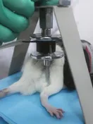
Figure 1 Tuina manipulation emulator.

Figure 2 Efects of tuina treatment on the solar-thermal pain threshold in a rat model of sciatic nerve injury.
Quantifcation of muscle atrophy
Gastrocnemius atrophy is a common symptom of sciatic nerve injury due to muscle denervation; the degree of muscle atrophy depends on injury severity and speed of nerve recovery (Wu et al., 2015; Tufaha et al., 2016). At 7 and 27 days aTher sciatic nerve injury, the recovery rate of muscle wet weight was evaluated in eight rats from each group. Briefy, fasting and water deprivation were performed for 24 hours prior to surgery. The rats were intraperitoneally anesthetized with a premixed solution containing 10% chloral hydrate (350 mg/kg body weight). The right lateral thigh area was shaved and the skin was disinfected with 10% povidone iodine. The right and leThgastrocnemius muscle was exposed through the crural-splitting approach. The recovery rate of muscle wet weight was defned by muscle weight of the experimental side divided by muscle weight of the control side (Jiang et al., 2016).
Morphological observation
At 7 and 27 days after sciatic nerve injury, morphology of the sciatic nerve was observed in eight rats from each group. After anesthesia with chloral hydrate and intracardial perfusion with normal saline, sciatic nerves were fxed with 4% paraformaldehyde in 0.1 M phosphate-buffered saline (pH 7.4) for 24 hours. The sciatic nerves were washed fully with water, dehydrated through a graded alcohol series, permeabilized with xylene, and embedded in parafn. The sciatic nerve was perpendicular to the long axis of the nerve transverse section, and then stained with hematoxylin and eosin. Morphology of the sciatic nerve was observed using a light microscope (Motic, Xiamen, Fujian Province, China).
Western blot assay
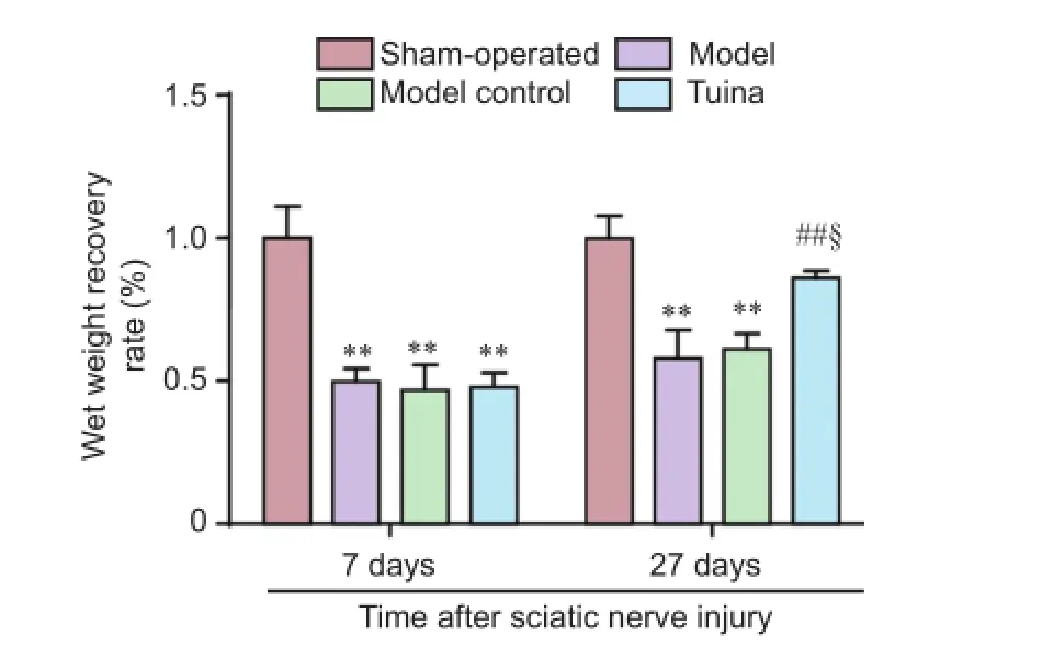
Figure 3 Efects of tuina treatment on the recovery rate of wet weight of gastrocnemius muscle in a rat model of sciatic nerve injury.
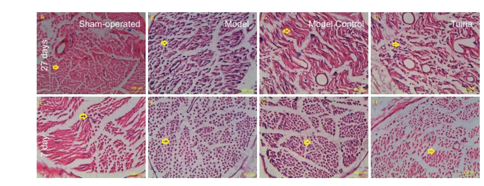
Figure 4 Efects of tuina treatment on morphology of injured sciatic nerves of rats at 7 days and 27 days aTher sciatic nerve injury (hematoxylin-eosin staining, × 400).
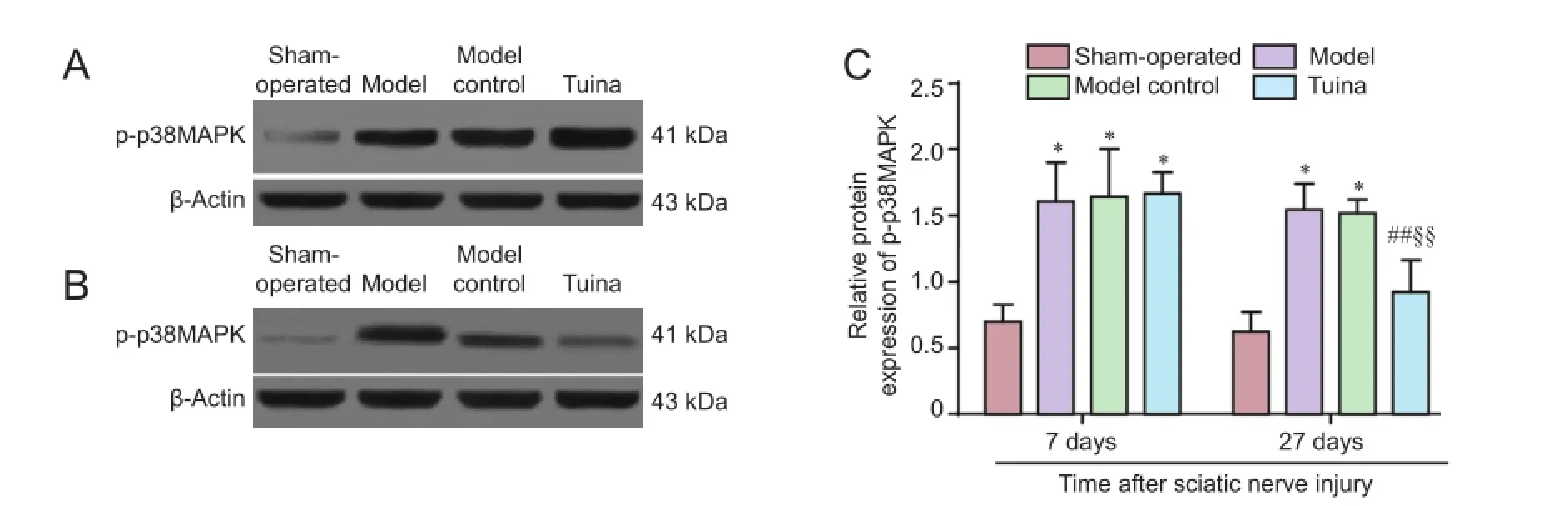
Figure 5 Efects of tuina treatment on p-p38MAPK in the dorsal horn of the spinal cord in a rat model of sciatic nerve injury.
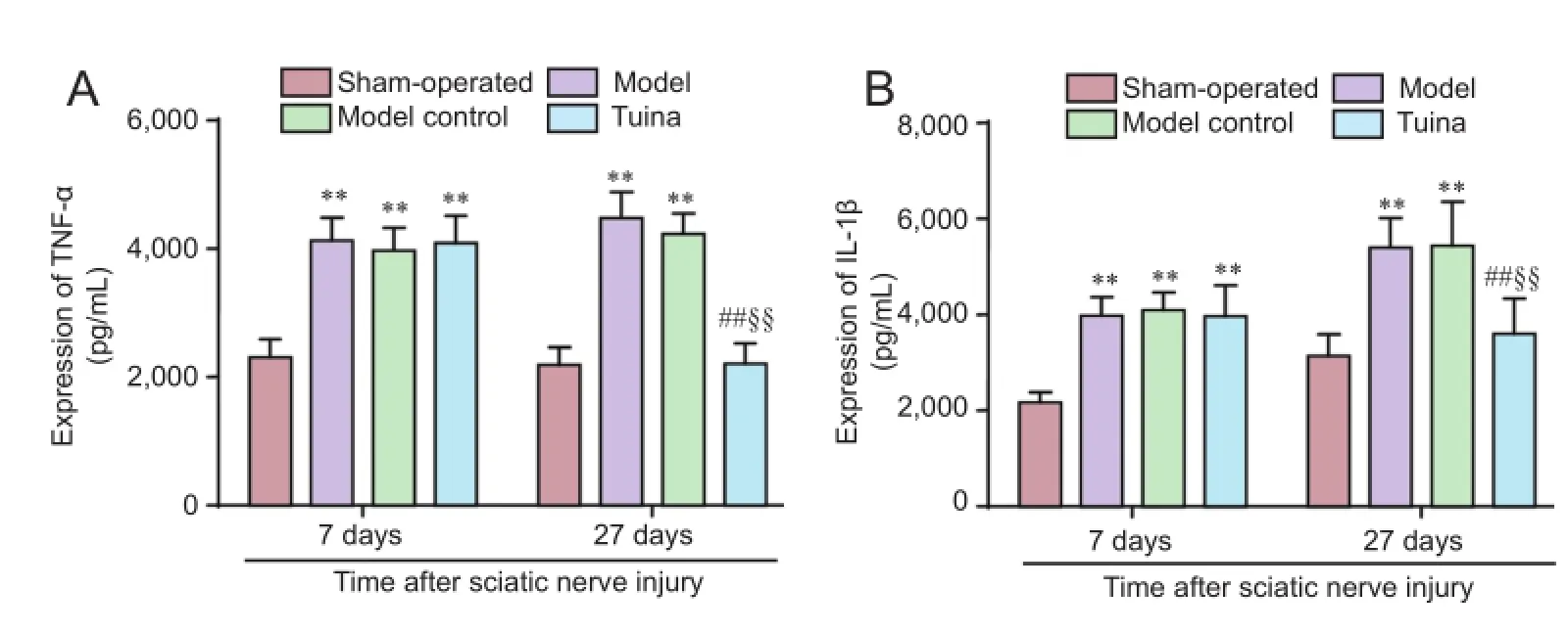
Figure 6 Efects of tuina treatment on TNF-α (A) and IL-1β (B) in the spinal cord of rats with injured sciatic nerves (enzyme-linked immunosorbent assay).
At 7 and 27 days after sciatic nerve injury, four rats from each group were used for western blot analysis of phospho-p38MAPK (p-p38MAPK) expression. The rats were anesthetized with chloral hydrate. ATher taking a blood sample from the abdominal aorta, the injured side of the lumbar spinal cord was extracted (L4-6) on an iced tray. The specimen was then placed in a tub of ice. The spinal cord was equally divided into two coronal parts. The inferior half of the spinal cord, namely the dorsal horn, was placed in liquid nitrogen for preservation (Shao et al., 2014). The different groups of cellular proteins were extracted, and the protein concentration was determined using the Coomassie brilliant blue assay. Each well was flled with 50 μg of protein sample. The samples were then separated on a sodium dodecyl sulfate-polyacrylamide gel by electrophoresis. Separated proteins in the gels were electrophoretically transferred onto polyvinylidene fuoride membranes at a constant voltage of 100 V at 4°C. The membranes were blocked in 6% skimmed milk powder for 2 hours, and then incubated with goat anti-rat p-p38MAPK monoclonal antibody (1:1,000; No. ab38238, Abcam, Cambridge, UK) and goat anti-rat β-actin monoclonal antibody (1:2,000; No. AC001-M, Santa Cruz Biotechnology, Santa, Cruz, CA, USA) at 4°C overnight. ATherwards, the membranes were washed with phosphate-buffered saline, and incubated with rabbit anti-goat horseradish peroxidase-IgG (1:5,000; No. SH-0031, Beijing, China) at room temperature for 1 hour. The membranes were then detected using enhanced chemiluminescence (ECL-0012, Pierce, FL, USA), X-ray exposure imaging was performed using a scanning analysis software system (Labworks? Analysis SoThware, ProteinSimple, Silicon Valley, CA, USA). β-Actin served as the standard reference.Relative protein expression levels were expressed as the integrated optical density ratio of each target protein to β-actin.
Enzyme-linked immunosorbent assay (ELISA)
At 7 and 27 days aTher sciatic nerve injury, four rats from each group were used for ELISA analysis of TNF-α and IL-1β protein expression in the spinal cord. ATher sacrifce by blood-letting through the abdominal aorta, the injured side of the lumbar spinal cord (L4-6) was extracted on an iced tray. The ELISA kit was used to detect expression levels of TNF-α (No. CSBE11987r, CUSABIO, Wuhan, Hubei Province, China) and IL-1β (No. CSB-E08055r, CUSABIO) in the L4-6spinal segment.
Statistical analysis
Data, expressed as the mean ± standard error of mean, were analyzed using SPSS 23.0 software (SPSS Inc, Chicago, IL, USA). One-way analysis of variance and post hoc least signifcant diference test were used to analyze data that obeyed normal distribution and homogeneity of variance. A value of P < 0.05 was considered statistically signifcant.
Results
Efects of tuina treatment on the solar-thermal pain threshold in a rat model of sciatic nerve injury
As shown in Figure 2, prior to intervention, a significant increase in paw withdrawal latency was detected in the sciatic nerves (P < 0.01, vs. sham-operated group). On day 20 post-intervention, the paw withdrawal latency of the tuina group was signifcantly decreased compared with the model (P < 0.01) and model control group (P < 0.05). Paw withdrawal latency of the tuina group was similar to the sham-operated group (P > 0.05).
Efects of tuina treatment on the gastrocnemius muscle atrophy in a rat model of sciatic nerve injury
As shown in Figure 3, prior to intervention, a signifcant decrease was found in the recovery rate of wet weight of gastrocnemius muscle subjected to sciatic nerve crush injury (P <0.01, vs. sham-operated group). On day 20 post-intervention, the recovery rate of wet weight of gastrocnemius muscle was signifcantly increased in the tuina group compared with the model and model control groups (P < 0.01).
Efects of tuina treatment on the morphology in a rat model of sciatic nerve injury
As shown in Figure 4, prior to intervention, the myelin sheath and axon were observed under a microscope in the sham-operated group; scattered myelin and axonal collapse were visible in the model, model control, and tuina groups. On day 20 post-intervention, the integrated structures of nerve fbers, axons, and Schwann cells were observed under a microscope in the sham-operated group. Disintegrated axis, broken myelin sheaths, and many Schwann cells were observed in the model and model control groups. The sciatic nerves were essentially normal, orderly, clear, and complete; nerve fiber axons and recovery of Schwann cells were observed in the tuina group.
Efects of tuina treatment on p-p38MAPK, TNF-α and IL-1β in in a rat model of sciatic nerve injury
The results of western blot assay showed a significant increase in p-p38MAPK in the model, model control, and tuina groups on day 0 post-intervention (P < 0.05, vs. sham-operated group). On day 20 post-intervention, p-p38MAPK values in the tuina group were significantly decreased compared with the model and model control groups (P <0.01), whereas values in the tuina group were similar to the sham-operated group (P > 0.05; Figure 5).
As shown in Figure 6, prior to intervention, a signifcant increase in TNF-α and IL-1β levels was detected in the sciatic nerves with sciatic nerve crush injury (P < 0.01, vs. sham-operated group). On day 20 post-intervention, TNF-α and IL-1β levels were significantly decreased in the tuina group compared with the model and model control groups (P< 0.01). These values were similar between the tuina group and the sham-operated group (P > 0.05).
Discussion
Recent studies have found that a phenotypic change in glial cells plays a key role in transmission of information along the entire nociceptive pathway. p38MAPK is mainly expressed in microglia in the dorsal horn of the spinal cord, and peripheral nerve injury can result in increased p38MAPK phosphorylation in the dorsal horn (Zhou et al., 2014b; Tatsumi et al., 2015). Previous studies of peripheral nerve injury treatment with tuina have shown that tuina regulates nerve growth factor and its related receptors in dorsal horn neurons and C fbers in the superfcial layers of the spinal cord to promote repair of peripheral nerve injury (Mei et al., 2013a). These fndings provide evidence that tuina regulates relevant regions and indices of the dorsal horn, as well as promotes recovery of sensory dysfunction in rats with injured sciatic nerve.
Sciatic nerve injury is a common standard and well-established peripheral nerve injury model to investigate the impact of different treatments in neural injury repair (Ma et al., 2013). This method is relatively inexpensive and easy to perform; the capacity for repairing this type of injury is equivalent in rats and subhuman primates (Marcolino et al., 2013). According to Chinese medicine, sciatic nerve injury is classified as a form of arthromyodynia, and is usually ascribed to meridian obstruction. Professor Yu innovated the “acupoint-nerve-muscle” theory based on many years of clinical practice and laboratory research, and this theory was used to select acupoints for the present study (Pan et al., 2015). Based on the “Three Methods and Three Points,”Yinmen, Weizhong, and Yanglingquan are located along three nerves: the sciatic, common peroneal nerve, and tibial nerve, respectively. The muscular locations of the three acupoints are the biceps femoris, semitendinosus, and anterior tibial muscle, respectively. Based on channel theory of traditional Chinese medicine, Yinmen and Weizhong belong to the Taiyang Bladder Foot Channel. Yanglingquan is located on the Shaoyang Gallbladder Foot Meridian. Therefore, the tuina manipulations based on the “Three Methods and ThreePoints” were designed to stimulate the anatomical structures of the meridians as a scientifc basis for treatment.
To further determine whether Chinese tuina has an impact on p38MAPK in microglia, we evaluated the infuence of Chinese tuina therapy on rehabilitation of sensory dysfunction post-sciatic nerve injury, specifcally p38MAPK phosphorylation and the inhibitory efect of tuina on phosphorylation of p38MAPK, TNF-α, and IL-1β in the dorsal horn. The primary fnding of the present study is that Chinese tuina signifcantly improved recovery of sensory dysfunction caused by peripheral nerve injury, as well as modifed expression of p-p38MAPK, TNF-α, and IL-1β in a rat model of sciatic nerve injury.
Since the introduction of the solar heat pain threshold measurement (Fruhstorfer et al., 1976), it has become a mainstay in recovery assessments of sensory function post-sciatic nerve injury. This study shows that tuina intervention of “Three Methods and Three Points” can improve paw withdrawal latency and the gastrocnemius muscle in the hind limbs of rats with an injured sciatic nerve, as well as contribute to nociceptive recovery and promote recovery of sensory dysfunction and muscle atrophy after peripheral nerve injury. Moreover, the “Three Methods and Three Points” can also promote morphological recovery of sciatic nerve fbers, Schwann cells, and axons, maintain the relative integrity of the sciatic nerve, promote recovery of sensory dysfunction after peripheral nerve injury, and provide behavioral recovery to a certain extent.
The regulation of p38MAPK in the dorsal horn of the spinal cord by endogenous substances or related treatments (Ostenfeld et al., 2013; Wang et al., 2016), can inhibit the phosphorylation of p38MAPK (Moon et al., 2013; Taves et al., 2016), and thereby afect TNF-α and IL-1β in the dorsal horn of the spinal cord (Kato et al., 2013; Berta et al., 2016). Inhibition of p38MAPK phosphorylation is therefore a possible therapeutic strategy to treat sensory dysfunction aTher peripheral nerve injury.
To further explore whether tuina influences p38MAPK phosphorylation, TNF-α, and IL-1β in the dorsal horn of the spinal cord, we performed tuina intervention based on the“Three Methods and Three Points” in sciatic nerve injury rats. In this study, p-p38MAPK expression increased in the sciatic nerve injury groups, as evidenced by increased levels on day 0 post-treatment. These results showed that p38MAPK activation in the dorsal horn led to increased p38MAPK phosphorylation in the dorsal horn of the spinal cord aTher sciatic nerve injury, thereby promoting production of infammatory factors and triggering increased TNF-α and IL-1β levels in the spinal cord. In addition to axonal disintegration and Schwann cell proliferation, p38MAPK was also involved in the formation and development of sensory dysfunction aTher peripheral nerve injury. Tuina treatment reduced expression of p-p38MAPK, TNF-α, and IL-1β, indicating decreased p38MAPK phosphorylation and production of infammatory factors. Tuina treatment using the “Three Methods and Three Points” induced recovery of sciatic nerve fibers, Schwann cells, and axons, and reduced peripheral nerve injury, thereby promoting recovery of sensory function.
The use of an animal model in the present study presented some notable limitations. The tuina treatment process could cause stress and panic in the rats, which could influence treatment efficacy. Additionally, owing to the individual differences in size and sensitivity between animal subjects, both the force and duration of each tuina manipulation was accordingly changed. The third limitation in the present study concerned the number of animals used in the study; p-p38MAPK, TNF-α, and IL-1β expression levels were only analyzed on day 0 and 20 post-sciatic nerve injury. Consequently, levels of p-p38MAPK, TNF-α and IL-1β at intermediary times were not measured, and it is therefore impossible to determine the intermediate rate of change.
In summary, p38MAPK in the spinal cord is involved in signal transduction of sensory dysfunction following peripheral nerve injury, which greatly infuences the production of TNF-α and IL-1β, which is central to hyperalgesia maintenance in a rat model of sciatic nerve injury. One of the mechanisms of tuina in treating peripheral nerve injury using the“Three Methods and Three Points” involves the regulation of p-p38MAPK expression in the dorsal horn, which subsequently inhibits TNF-α and IL-1β expression and promotes recovery of behavior and morphology aTher peripheral nerve injury. These results provide a scientific basis for the use of tuina in the clinical treatment of peripheral nerve injury.
Author contributions:XG participated in design and performance of the main experiments. TYY was responsible for study design and guidance. SW modifed language throughout the entire article. CM and YHT took charge of functional evaluation. SW and CY took charge of immunohistochemistry. JLL and TTL were responsible for perfusion and tissue processing. MQL provided the instruction for modeling. WDJ helped with data analysis. All authors approved the fnal version of the paper.
Conficts of interest:None declared.
Plagiarism check:This paper was screened twice using CrossCheck to verify originality before publication.
Peer review:This paper was double-blinded and stringently reviewed by international expert reviewers.
Berta T, Qadri YJ, Chen G, Ji RR (2016) Microglial signaling in chronic pain with a special focus on caspase 6, p38 MAP kinase, and sex dependence. J Dent Res 95:1124-1131.
Bozkurt A, Boecker A, Tank J, Altinova H, Deumens R, Dabhi C, Tolba R, Weis J, Brook GA, Pallua N, van Neerven SG (2016) Efcient bridging of 20 mm rat sciatic nerve lesions with a longitudinally micro-structured collagen scafold. Biomaterials 75:112-122.
Chen JF, Shu XN, Tang SJ, Wu Y, Zhang YP (2016) Infuence of lumbar disc degeneration on the efcacy of lumbar fxed-point rotation manipulation in sitting position: a fnite element study. J Acupunct Tuina Sci 14:295-299.
Chowdhry S, Davis J, Boyd T, Choo J, Brooks RM, Kelishadi SS, Tutela JP, Yonick D, Wilhelmi BJ (2015) Safe tummy tuck: anatomy and strategy to avoid injury to the lateral femoral cutaneous nerve during abdominoplasty. Eplasty 15:e22.
Deng DW, Lin JP, Chen SQ, Xia M, Wang SZ (2016) Effects of electro-acupuncture of “Weizhong” point on the expressions of caspase3 and XIAP in rat with lumbar disc degeneration. Zhongguo Kangfu Yixue Zazhi 31:394-398.
Fruhstorfer H, Lindblom U, Schmidt WC (1976) Method for quantitative estimation of thermal thresholds in patients. J Neurol Neurosurg Psychiatry 39:1071-1075.
Gao YF, Wu JC, Geng N, Yu TY (2013) Effect of Tuina therapy on motor function and scafolding proteins NF-M in neurons of sciatic nerve injury rat models. Huanqiu Zhongyiyao 6:894-897.
Gao YF, Yao BB, Wu JC, Wen M, Yu Y, Yu TY (2014) Effect of massage on MAP-2 and NF-M expression of sciatic nerve injury in rats. Zhongyiyao Daobao 20:11-13.
Goswami R, Anastakis DJ, Katz J, Davis KD (2016) A longitudinal study of pain, personality, and brain plasticity following peripheral nerve injury. Pain 157:729-739.
Hu CN, Wang SH (2016) Clinical observation on oblique Ban-pulling tuina manipulation plus acupuncture for cervical vertigo. J Acupunct Tuina Sci 14:26-30.
Hu L, Zhang HL, Tu Y, Lu J (2012) The research progress of alleviating exercise-induced fatigue by acupuncture-moxibustion and tuina during the past fve years. Zhenjiu Linchuang Zazhi 28:69-71.
Jang HJ, Kim JY, Han JD, Lee HJ, Kim JS, Park JS, Choi RK, Choi YJ, Shim WH, Kwon SW, Kim TH (2016) Numbness after transradial cardiac catheterization: the results from a nerve conduction study of the superfcial radial nerve. Korean Circ J 46:161-168.
Jiang W, Wang Y, Tang J, Peng J, Wang Y, Guo Q, Guo Z, Li P, Xiao B, Zhang J (2016) Low-intensity pulsed ultrasound treatment improved the rate of autograft peripheral nerve regeneration in rat. Sci Rep 6:22773.
Kato N, Matsumoto M, Kogawa M, Atkins GJ, Findlay DM, Fujikawa T, Oda H, Ogata M (2013) Critical role of p38 MAPK for regeneration of the sciatic nerve following crush injury in vivo. J Neuroinfammation 10:1.
Lu MQ (2016) Efect of Tuina on growth oriented mechanism of axon growth in nerve repair and regeneration of SNI rats. Beijing: Beijing University of Chinese Medicine.
Lu MQ, Yu TY, Yao BB, Chen GY, Pan F, Xian ST, Wu JC, Mao YQ, Zhang LF (2015) Efects of tuina therapy on the neural ultrastructure of sciatic nerve injury model rats. Nanjing Zhongyiyao Daxue Xuebao 31:349-352.
Ma J, Liu J, Yu H, Wang Q, Chen Y, Xiang L (2013) Curcumin promotes nerve regeneration and functional recovery in rat model of nerve crush injury. Neurosci Lett 547:26-31.
Marcolino AM, Barbosa RI, das Neves LMS, Mazzer N, de Jesus Guirro RR, de Cássia Registro Fonseca M (2013) Assessment of functional recovery of sciatic nerve in rats submitted to low-level laser therapy with different fluences. An experimental study: laser in functional recovery in rats. J Hand Microsurg 5:49-53.
Mei XH, Ji Q, Yao BB, Wu JC, Lu MQ, Yu TY (2013a) Investigation of tuina therapy on NGF and p75NTR of sciatic nerve injury model rats. Zhonghua Zhongyiyao Zazhi 28:1994-1997.
Mei XH, Ji Q, Wu JC, Pan F, Wang L, Yu TY (2013b) Infuences of tuina therapy on nerve growth factor and TrkA receptor of NGF in rats with sciatic nerve injury. Beijing Zhongyiyao Daxue Xuebao 36:497-500.
Moon JY, Roh DH, Yoon SY, Kang SY, Choi SR, Kwon SG, Choi HS, Han HJ, Beitz AJ, Lee JH (2013) Sigma-1 receptor-mediated increase in spinal p38 MAPK phosphorylation leads to the induction of mechanical allodynia in mice and neuropathic rats. Exp Neurol 247:383-391.
Nishihara T, Remacle AG, Angert M, Shubayev I, Shiryaev SA, Liu H, Dolkas J, Chernov AV, Strongin AY, Shubayev VI (2015) Matrix metalloproteinase-14 both sheds cell surface neuronal glial antigen 2 (NG2) proteoglycan on macrophages and governs the response to peripheral nerve injury. The Journal of Biological Chemistry 290:3693-3707.
Ostenfeld T, Krishen A, Lai RY, Bullman J, Baines AJ, Green J, Anand P, Kelly M (2013) Analgesic efcacy and safety of the novel p38 MAP kinase inhibitor, losmapimod, in patients with neuropathic pain following peripheral nerve injury: a double-blind, placebo-controlled study. Eur J Pain 17:844-857.
Pan F, Yu TY, Wong S, Xian ST, Lu MQ, Wu JC, Gao YF, Li XQ, Geng N, Yao BB (2015) Chinese tuina downregulates the elevated levels of tissue plasminogen activator in sciatic nerve injured Sprague-Dawley rats. Chin J Integr Med doi: 10.1007/s11655-015-2142-1.
Shao YJ, Yan M, Zhang H, Zhao W, Li DF (2014) Efects of curcumin on expression of PDCD5 of spinal dorsal horn in rats with chronic construction injury on sciatic nerve. Huanan Guofang Yixue Zazhi 28:417-423.
Shen ZF, Bian XD, Gao F, Li QJ, Yuan JY (2015) Efect of tuina manipulations on blood pressure and its variability in hypertension patients. J Acupunct Tuina Sci 13:180-184.
Sunderland S (1951) A classifcation of peripheral nerve injuries producing loss of function. Brain 74:491-516.
Tatsumi E, Yamanaka H, Kobayashi K, Yagi H, Sakagami M, Noguchi K (2015) RhoA/ROCK pathway mediates p38 MAPK activation and morphological changes downstream of P2Y12/13 receptors in spinal microglia in neuropathic pain. Glia 63:216-228.
Taves S, Berta T, Liu DL, Gan S, Chen G, Kim YH, Van de Ven T, Laufer S, Ji RR (2016) Spinal inhibition of p38 MAP kinase reduces inflammatory and neuropathic pain in male but not female mice: Sex-dependent microglial signaling in the spinal cord. Brain, Behav, Immun 55:70-81.
Tuffaha S, Budihardjo J, Sarhane K, Khosheim M, Song D, Broyles J, Salvatori R, Means K, Higgins J, Shores J, Hoke A, Cooney D, Lee WPA, Brandacher G (2015) Abstract 129: Efects of growth hormone therapy on axonal regeneration, muscle atrophy, Schwann cell proliferation and end-organ reinnervation following nerve injury and repair. Plast Reconstr Surg 135:92-93.
Tufaha SH, Budihardjo JD, Sarhane KA, Khusheim M, Song D, Broyles JM, Salvatori R, Means KRJ, Higgins JP, Shores JT, Cooney DS, Hoke A, Lee WP, Brandacher G (2016) Growth hormone therapy accelerates axonal regeneration, promotes motor reinnervation, and reduces muscle atrophy following peripheral nerve injury. Plast Reconstr Surg 137:1771-1780.
Wang J, Zhang CK, Zhang Q, Yang F, Zhang T, Dong YL, Li JL (2016) Mechanism of triptolide (T10) exerting analgesic efect via inhibition of the phosphorylation of p38-MAPK in spinal dorsal horn of rats. Shenjing Jiepou Xue Zazhi 32:18-24.
Wang Y, Yang HY (2015) Review of the research on the mechanism of weizhong point in the treatment of lumbar-back pain. Zhenjiu Linchuang Zazhi 31:90-92.
Wen XG, Yan YJ, Wu YL (2015) Intensive stimulation tuina at tender points plus medication for cervical intervertebral disc herniation. J Acupunct Tuina Sci 13:377-380.
Wu F, Zhang B, Zheng HM, Wei JB, Zhang XH, Yu TY (2013) Behavior study of tuina treatment on sciatic nerve injury in rats. Chenghdu Zhongyiyao Daxue Xuebao 36:41-43.
Wu JC (2014) Efect of tuina on the recovery mechanism of SNI in rats by afecting the expression of TrkC and NT-3. Beijing: Beijing University of Chinese Medicine.
Wu R, Yan Y, Yao J, Liu Y, Zhao J, Liu M (2015) Calpain 3 expression pattern during gastrocnemius muscle atrophy and regeneration following sciatic nerve injury in rats. Int J Mol Sci 16:26927-26935.
Xu L, Yu XR, Huang YG (2012) P38 MAPK inhibitor decreases the TNF-α level in spinal cord of rats with chronic constriction Injury. Jichu Yixue yu Linchuang 32:1126-1131.
Xu X, Wang HY, Zhang ZW, Han H, Wang Y (2016) Efect of massage therapy on pulmonary functions of pediatric asthma: A systematic review and meta-analysis of randomized controlled trials. Eur J Integr Med 8:98-105.
Xu YS (2013) Construction of modern massage disciplines. Shiyong Zhongyi Neike Zazhi 27:152-153.
Yao BB, Mei XH, Wu JC, Yu TY (2013) Study on the infuence of massage to sciatic nerve injured rat’s axoplasmic transport function based on motor protein. Nanjing Zhongyiyao Daxue Xuebao 29:338-341.
Yu TY (2015) Massage. Beijing: China Traditional Medicine Press.
Zhao Z (2014) Preliminary study of function of the spinal microglial P2Y12 receptor and P2X7 receptor in activation of microglia in neuropathic pain. Chongqing: Third Military Medical University.
Zhong Y, Huang YL, XU JD (2012) Role of interleukin-1β in spinal LTP in rats with neuropathic pain. Zhongguo Bingli Shengli Zazhi 28:541-545.
Zhou C, Shi X, Huang H, Zhu Y, Wu Y (2014a) Montelukast attenuates neuropathic pain through inhibiting p38 mitogen-activated protein kinase and nuclear factor-kappa B in a rat model of chronic constriction injury. Anesth Analg 118:1090-1096.
Zhou TT, Wu JR, Chen ZY, Liu ZX, Miao B (2014b) Efects of dexmedetomidine on P2X4Rs, p38-MAPK and BDNF in spinal microglia in rats with spared nerve injury. Brain Res 1568:21-30.
Copyedited by Cooper C, de Souza M, Yu J, Li CH, Qiu Y, Song LP, Zhao M
*Correspondence to: Tian-yuan Yu, Ph.D., yutianyuan@sina.com.
orcid:
0000-0002-5455-5833 (Xin Guo)
0000-0003-2089-3101 (Tian-yuan Yu)
10.4103/1673-5374.197147
Accepted: 2016-11-24
 中國(guó)神經(jīng)再生研究(英文版)2016年12期
- 中國(guó)神經(jīng)再生研究(英文版)的其它文章
- Expression changes of nerve cell adhesion molecules L1 and semaphorin 3A aTher peripheral nerve injury
- Injury of the arcuate fasciculus in a patient with progressive bulbar palsy
- Biodegradable magnesium wire promotes regeneration of compressed sciatic nerves
- Electroacupuncture at Dazhui (GV14) and Mingmen (GV4) protects against spinal cord injury: the role of the Wnt/β-catenin signaling pathway
- Application of a paraplegic gait orthosis in thoracolumbar spinal cord injury
- Fine motor skill training enhances functional plasticity of the corticospinal tract aTher spinal cord injury