
Role of neurotrophic factors in enhancing linear axonal growth of ganglionic sensory neurons in vitro
2020-03-07 05:31:26MicheleFornaroAlessiaGiovannelliAngelicaFoggettiLuisaMuratoriStefanoGeunaGiorgiaNovajraIsabellePerroteau
中國神經(jīng)再生研究(英文版) 2020年9期
Michele Fornaro , Alessia Giovannelli, Angelica Foggetti, Luisa Muratori, , Stefano Geuna, , Giorgia Novajra, Isabelle Perroteau,
1 Department of Anatomy, College of Graduates Studies (CGS), Chicago College of Osteopathic Medicine (CCOM), Midwestern University, Downers Grove, IL, USA
2 Department of Clinical and Biological Sciences, University of Turin, Torino, Italy
3 Institute of Physiology, Christian-Albrechts-University Kiel, Kiel, Germany
4 Neuroscience Institute Cavalieri Ottolenghi (NICO), Torino, Italy
5 Department of Applied Science and Technology, Politecnico di Torino, Torino, Italy
Abstract Neurotrophins play a major role in the regulation of neuronal growth such as neurite sprouting or regeneration in response to nerve injuries. The role of nerve growth factor, neurotrophin-3, and brain-derived neurotrophic factor in maintaining the survival of peripheral neurons remains poorly understood. In regenerative medicine, different modalities have been investigated for the delivery of growth factors to the injured neurons, in search of a suitable system for clinical applications. This study was to investigate the inf luence of nerve growth factor, neurotrophin-3 and brain-derived neurotrophic factor on the growth of neurites using two in vitro models of dorsal root ganglia explants and dorsal root ganglia-derived primary cell dissociated cultures. Quantitative data showed that the total neurite length and tortuosity were differently inf luenced by trophic factors. Nerve growth factor and, indirectly, brain-derived neurotrophic factor stimulate the tortuous growth of sensory fibers and the formation of cell clusters. Neurotrophin-3, however, enhances neurite growth in terms of length and linearity allowing for a more organized and directed axonal elongation towards a peripheral target compared to the other growth factors. These findings could be of considerable importance for any clinical application of neurotrophic factors in peripheral nerve regeneration. Ethical approval was obtained from the Regione Piemonte Animal Ethics Committee ASLTO1 (file # 864/2016-PR) on September 14, 2016.
Key Words: brain-derived neurotrophic factor; directionality; dorsal root ganglia explant; nerve growth factor; nerve regeneration; neurite growth enhancement; neurotrophic factors; neurotrophin-3; sensory neurons; tortuosity
Introduction
After injury, peripheral nerve repair spontaneously occurs via growth cones located at the tips of axons. A variety of stimuli from the surrounding environment can be detected by these growth cones. The subsequent response to these cues results in axonal outgrowth, possibly over distances of hundreds of microns to reconnect to the end-tissue target (Miller et al., 2001; Neto et al., 2017; Sulaiman et al., 2018; Kornfeld et al., 2019). In some cases, however, there may be a relatively large gap between the severed nerve ends and regenerating axon sprouts fail to reach their end target due to a lack of directionality. The failure of the regenerating axons to quickly re-establish contact with target tissue leads to permanent loss of sensory or motor function. A better understanding of the mechanisms that can guide and direct axons along a desired path are critical for future clinical applications.
Neurotrophic factors (NTs) play a critical role in neuronal survival after nerve injury by protecting neurons from death, thus increasing the potential for axon regeneration. While postnatal and adult sensory neurons do not require NTs for their continued survival, the maintenance of mature phenotypic characteristics and physiologic responsiveness seems to be dependent upon NTs such as nerve growth factor (NGF) and neurotrophin-3 (NT-3) (Kimpinski et al., 1997; Mamounas et al., 2000; Yamamoto and Hanamura, 2005; Santos et al., 2016; Cacialli et al., 2019). Although NGF, NT-3, and brain-derived neurotrophic factor (BDNF) have been described as important factors to promote axonal regrowth through a conserved cell polarity-signalling pathway (Grider et al., 2005; Bagheri et al., 2019; Katebi et al., 2019; Melo et al., 2019), application of these factors in regenerative medicine has seen very poor results thus far (Mitchell et al., 2016). In particular, most of unsuccessful experiments implied a topical application of NTs at the site of lesion. Axons were definitively incentivized to regrow as an unorganized bundle of fibers with no directionality and unable or barely able to reach the periphery, thus impeding functional motor or sensory recovery. At the site of lesion, NGF was found to generate and maintain hypersensitivity by inducing aberrant sprouting and/or neuroma formation in response to tissue and/or nerve injury (Mantyh et al., 2011).
Therefore, the purpose of this study was to investigate the activity of the NT family on neurite growth in vitro with the overall goal of identifying a key player for an organized and oriented fiber regrowth, thus opening possible applications in the reconstructive and regenerative medicine procedures.
The singular effects of NGF, BDNF and NT-3 were studied in two in vitro models: 1) a co-culture of DRG-derived dissociated neuronal and glial cells; 2) dorsal root ganglia (DRG) explants. The effect of BDNF was also tested on sciatic nerve-derived primary culture of Schwann cells.
Materials and Methods
Animals
Adult female Sprague-Dawley rats (Harlan, Barcelona, Spain), weighing 175-200 g, were used in this study. Animals were housed in large cages in a temperature and humidity controlled room with 12-hour light/dark cycles. The animals were fed with standard chow and water ad libitum. Adequate measures were taken to minimize pain and discomfort taking into account humane endpoint criteria for animal suffering and distress. All procedures were performed in accordance with the European Communities Council Directive of November 24, 1986 (86/609/EEC) and approved by the Regione Piemonte Animal Ethics Committee ASLTO1 (file # 864/2016-PR) on September 14, 2016.
DRG explants
Rats were sacrificed by a lethal intramuscular injection of tiletamine + zoletil (3 mg/kg, Virbac, Carros, France) in the back thigh. The vertebral column was surgically dissected and the vertebral body was removed to gain a ventral access to the spinal cord. DRG from all spinal levels were localized, removed, and isolated. The dissecting procedure was completed within 90 minutes. DRG were collected in a 3.5 mL Petri dish with 1.8 mL of F12 medium (Gibco, Carlsbad, CA, USA). The connective-tissue capsules surrounding the ganglia were reduced with f ine forceps. The ganglia were adhered onto matrigel-coated coverslips (BD Biosciences, Bedford, MA, USA) and incubated at 37°C for 1 hour (Fornaro et al., 2018). Matrigel was diluted 1:1 in serum- free medium (SFM) (Fornaro et al., 2018). Explants were maintained at 37°C with 5% CO2.
DRG primary culture
DRG were harvested, collected, and then enzymatically dissociated in F12 medium containing 12.5 mg/mL collagenase type IV (Sigma, St. Louis, MO, USA) for 1 hour (37°C, 5% CO2). The incubation in collagenase was repeated for 45 minutes.
DRGs were washed three times with F12 and incubated in bovine pancreatic trypsin 2 × 25 mg/mL (Sigma) for 30 minutes (37°C, 5% CO2). The trypsin was inactivated with 33% fetal bovine serum (FBS; VWR, Radnor, PA, USA) in F12 medium. The medium was removed and DRG were washed three times with F12 medium. Then the ganglia were transferred to a 15 mL falcon with 2 mL F12 medium and mechanically dissociated by gentle trituration using a glass Pasteur pipette. Dissociated cells were f iltered through a 70 μm-pore sized filter (BD Biosciences, San Jose, CA, USA), transferred to a 15 mL falcon, and then centrifuged at 25 × g for 5 minutes. Pellets were re-suspended in 0.5 mL of supernatant and 1 mL of F12 medium containing 0.5 mL of F12 and 0.5 mL of 30% bovine serum albumin (Sigma). A differential centrifugation with bovine serum albumen at 75 × g for 10 minutes was employed to separate neurons and a small amount of satellite cells from most of Schwann cells, astrocytes and connective tissue of the ganglionic capsules. The supernatant was completely removed and the pellet containing neurons and satellite cells were re-suspended in F12 medium. The pellet containing Schwann cells and f ibroblasts was discarded.
Dissociated neurons and satellite cells were seeded on glass coverslips pre-coated with laminin (2 μg/cm2, Sigma) in 24-well culture dishes with SFM and incubated at 5% CO2at 37°C for 24, 48 and 72 hours. The primary culture of glial cells with a high representation of satellite cells was maintained in vitro for several passages.
Schwann cell primary culture
Primary Schwann cells cultures were obtained from sciatic nerves harvested from adult Sprague Dawley rats. The nerves were collected in cold DMEM plus glutamax (Gibco, Carlsbad, CA, USA) containing 100 U/mL penicillin (R&D system, Minneapolis, MN, USA) and 100 g/mL Streptomycin (R&D system, Minneapolis, MN, USA). Nerves were then gently dissected and the epineurium removed with f ine forceps. Fragments of nerves were then plated in 35 mm petri dishes and incubated with glial permissive medium (DMEM plus glutamax, 100 U/mL penicillin and 100 g/mL streptomycin, 14M forskolin and 100 ng/mL LNRG11; R&D System, Minneapolis, MN, USA) for 2 weeks under culture conditions (37°C, 5% CO2). The medium was replenished every 72 hours. The culture medium was then enriched with 0.125% collagenase type IV (ThermoFisher, Lafayette, CO, USA) and 117 U/mg dispase (Gibco) for 24 hours and mechanically dissociated with a Pasteur glass pipette. The cell suspension was f iltered through a 70 μm cell strainer (BD Biosciences) and centrifuged at 100 × g for 5 minutes. The cell pellet was resuspended in glial permissive medium, seeded on 35 mm petri dishes pre-coated with poly-D-lysine (Sigma) and incubated under culture conditions. An antibody complement method was adopted to purify Schwann cells from f ibroblasts (Tohill et al., 2004; Kaewkhaw et al., 2012; Pascal et al., 2014).
NT treatments
To evaluate the direct effects of NTs on neurite outgrowth, primary cultures and DRG explants were treated with SFM with or without neurotrophic factors NGF (50 ng/mL, 2.5S beta-NGF, Sigma) (Malcangio et al., 1997; Turney et al., 2016; Naletova et al., 2019), BDNF (10 ng/mL, Sigma) (Santos et al., 2016) and NT-3 (10 ng/mL, Sigma) (Malcangio et al., 1997; Turney et al., 2016) for 24, 48 and 72 hours. To test for the lack of dependence on these neurotrophic factors neutralizing antibody against NGF (1 μg/mL, Sigma), BDNF (1 μg/mL, Sigma) and NT-3 (1 μg/mL, Sigma) were added to culture medium. To evaluate whether BDNF specif ically activates satellite cells or both satellite and Schwann cells, the two glial populations were cultured in Dulbecco’s Modif ied Eagle Medium (DMEM) (1 g/L glucose), 10% FBS, 1% penicillin/streptomycin with 63 ng/mL of glial growth factor and 10 μM of forskolin with or without BDNF. The cultures were maintained in a humidif ied incubator at 37°C and 5% CO2for 72 hours. The cell cultures were then processed for immunof luorescence.
Immunocytochemistry
To evaluate the morphology and neurite outgrowth, cell cultures and explants were f ixed with 4% paraformaldehyde for 20 minutes. After rinsing with phosphate buffered saline (PBS pH 7.2), the cells were incubated in 0.1% triton X-100, 10% normal goat serum (NGS, Thermo Scientific, Bedford, MA, USA)/0.1% NaN3, for 1 hour. Samples were incubated for 1 hour at room temperature in primary antibodies and processed for single or double immunof luorescence. The primary antibodies used in this study were: mouse monoclonal anti-βIII Tubulin (1:1000, Thermo Scientific), rabbit polyclonal anti-S-100 (1:600, Abcam, Cambridge, MA, USA) and rabbit polyclonal anti-Peripherin (1:1000, Millipore, Temecula, CA, USA). After rinsing with PBS, the specimens were incubated for 1 hour at room temperature with secondary antibodies Alexa Fluor 488 anti-mouse IgG (1:200, Molecular Probes, Eugene, Oregon, USA) and Cy3 anti-rabbit IgG (1:200, Jackson Immunoresearch, West Grove, PA, USA). Finally, samples were mounted with a f luorescent mounting medium (Dako, carpinteria, CA, USA), imaged using the Nikon Eclipse Ti A1R confocal microscope (Nikon, Melville, NY, USA), and analyzed using the NIS-Elements software (Nikon).
Quantitative analysis of neurite length and tortuosity
Confocal images of dissociated cells cultures were analyzed with the Leica image-pro Premier software (Leika Microsystems, Buffalo Grove, IL, USA). The image 3D-mode was utilized to better resolve the fibers. The same software was utilized to measure the neurite length (L) and total distance (vector, v) from the tip of the neurite to its origin at the cell body. The GraphPad Prism 8.2.1 software (GraphPad Software, San Diego, CA, USA) was utilized for statistical analysis and graph preparation.
Confocal images of DRG explants were processed by image analysis software (ImageJ, NIH, Bethesda, MD, USA) to evaluate the spatial distribution and extension of neurites, using a modif ied Sholl analysis (Sholl, 1953). In Sholl analysis, the number of dendrite branches of a neuron is represented as a function of the distance from the cell body, by counting the number of intersections of neuritic processes with concentric circles centered in the cell soma. In this study, the perimeter of the ganglion was manually drawn on the image (Polygon selection tool) and then progressively enlarged (Edit-Selection-Enlarge tool) to create concentric curves at f ixed distance (7 μm) between each other, which were used as coordinates of reference for neurite analysis. The number of intersections of neuritic processes (both primary neurites and branches) with the different curves of DRG grown in the presence of each factor was blindly counted and represented in terms of mean values and standard deviation as a function of the distances from the ganglion perimeter.
For DRG explants treated with NGF and NT-3 (explants treated with BDNF did not differ significantly from control), primary neurites were traced on the image (segmented line tool). Their length (L) and the distance (d) between the neurite termination and its starting point from the ganglion were measured (Analyze-Measure tool). The neurite tortuosity (t) was calculated using the following formula:
The neurite tortuosity was reported as mean values and standard deviation. One-way analysis of variance was conducted to assess statistical significance.
Results
Neurotrophic effect on DRG-derived dissociated cells culture
Semi-purified co-culture of sensory neurons and satellite cells was seeded in vitro in a 2-dimensional system. After 48-72 hours in SFM, cells and their projections spontaneously formed clusters (Figure 1A-C). To study the effect of NTs on axonal regrowth, each singular factor was added to the culture medium at day-in-vitro (div) 0 and the effect of specif ic NT on cell morphology (both neuron and glia), cellcell interaction, axonal growth and linearity/tortuosity of the projections was analyzed. To assess the effect of each single factor, antibodies against the other two factors were added to the culture medium. Moreover, the effect of the single factor was null when added together with the antibody against the factor itself (negative control). DRG dissociated cells in the presence of NGF (Figure 1A1-C1) have a high tendency to cluster together, anticipating the normal aggregations of cells observed in SFM (Figure 1A-C). In the presence of NGF, a round, well-defined cluster of β-tubulin-positive neurons and S-100-positive glial cells formed in half of the time (24 hours instead of 48 hours). At 72 hours, the double immunocytochemistry showed a complex bundle of β-tubulin-positive neurites surrounding and def ining the cluster of neuronal and S-100-positive glial cells. These neurites did not leave the cluster or barely elongate outside of it. The cell aggregation was inhibited by adding anti-NGF to the culture medium (Figure 1C1 inset), confirming that NGF plays a role in favoring the cell-cell interaction and cluster formation. A cluster formation was observed also when cells were grown in the presence of NGF, anti-BDNF, and anti-NT-3 alone and in combination.
Similarly to what was seen for NGF, also in the presence of BDNF, cluster formation and presence of tortuous fibers were observed. However, the aggregation was delayed (72 hours) compared to cultures treated with NGF. The neurites of sensory neurons did not elongate far out from the roundshaped cluster (Figure 2A). No cell aggregation was observed when anti-BDNF antibody was added to the culture medium (data not shown). Interestingly, adding anti-NGF antibody to the cultures treated with BDNF prevented cell cluster formation (Figure 2B), while the same effect was not obtained after adding anti-NT-3 antibody (data not shown). The effect of BDNF was further evaluated in two different primary cultures of glial cells: DRG-derived satellite cells (Figure 2C and D) and sciatic nerve-derived Schwann cells (Figure 2E and F). BDNF-dependent morphological changes of the two cell populations were observed in confocal microscopy after cells were immunolabelled with the glial marker S-100. More significant morphological changes were seen in the satellite cell population when BDNF was added to the culture medium (Figure 2C and its inset). Cells looked more conf luent, possibly more numerous, bigger in size and displayed more and well developed processes branching out of the cell body compared to the same population grown in the presence of BDNF and anti-BDNF antibody (Figure 2D and its inset). On the other hand, no morphological changes were detected in sciatic nerve-derived Schwann cells culture in the presence of BDNF (Figure 2E) compared to cells grown with BDNF plus anti-BDNF antibody (Figure 2F), suggesting that BDNF may specif ically induce morphological changes in satellite cells but not in Schwann cells.
Contrary to the other two factors, when NT-3 was singularly added to the medium (in combination with anti-NGF and anti-BDNF antibodies) of DRG-derived cell cultures, no cell-cell aggregation was observed. Instead, NT-3 elicited more linear growth of neuronal processes (Figure 3). The effect of NT-3 was reverted when anti-NT-3 was added to the medium, allowing the formation of cell cluster and confused bundle of neuritis (Figure 3 inset).
The effect of each factor on neurite growth was evaluated in dissociated sensory neurons (Figure 4). Confocal images of β-tubulin and S-100 double immunolabelled cells were imported to the imaging software Leica Image-Pro Premiere and converted in 3D view to obtain a better resolution of the neurites (Figure 4A-C). The two parameters blindly measured were: (1) neurite’s length (L) and (2) linear growth resulting from the vector distance (V) between the cell body and the distal tip of the process (Figure 4D). A comparative analysis of the neurites grow in the presence of the growth factors showed a significant increase in the length of neurites in the presence of either NT-3 or NGF compared to in the presence of BDNF, and there was no significant statistical difference between the NT-3 and NGF groups (Figure 4E). However, analysis of the vector lengths showed a significant increase in the linear extension of neurites treated with NT-3 compared to that treated with NGF or BDNF, and there was no significant difference in linear extension of neurites between the NGF and BDNF groups (Figure 4F).
Neurotrophic effect on DRG explants
The activity of the NTs was also studied in DRG explants (Figure 5). The data obtained in the explants supported and confirmed the results shown in dissociated cells cultures. For a clear resolution of the neurite sprouts, all the measures were conducted on DRG grown for 3 days ex vivo (Figure 5A-C). A more obvious linear growth in the presence of NT-3 (Figure 5D) compared to NGF (Figure 5E) can be seen often 7 days in vitro. However, in this study, the effect of NGF, BDNF and NT-3 on axonal growth was quantif ied in terms of length and linearity/tortuosity of 3 days in vitro. Images of DRG explants treated with a singular growth factor were imported and analyzed and over-imposed with a grid of concentrical lines to quantify the total number of intersections between the grid and the neurite (Figure 5F). Moreover, length of neurites (L), and vector distance (d) were measured (Figure 5G). A quantitative analysis showed a significant increase in tortuosity (t = d/L) of neurites grown in the presence of NGF compared to NT-3, suggesting that NT-3 allows for a more linear growth (Figure 5H).
A different behavior of growing fibers in the presence of different factors is also shown in terms of the number of intersections with over-imposed grid counted for each condition (Figure 5I). In the presence of NGF, fibers wound around and have a higher percentage of intersections compared to fibers in the presence of NT-3 which grew more linearly and reached longer distances. Finally, explants treated with BDNF were not different from control (SFM) and showed a limited neurite growth.
Discussion
This study compared the contribution of NT family members in the regeneration of peripheral fibers in in vitro DRG models. This comparison highlights not only the different behavior of growing fibers in the presence of each single factor but also which factor is the key to facilitating a more organized and oriented neurite elongation under the regenerative medicine principle that a more linear and guided axonal growth results in a more effective nerve regeneration and increased function recovery after peripheral nerve lesion.
In this study, NGF, BDNF, and NT-3 were tested on two different in vitro models: DRG-derived dissociated primary cell cultures and DRG organotypic cultures. BDNF was also tested on the primary culture of Schwann cells derived from rat sciatic nerves. In serum-free medium condition, dissociated DRG cells spontaneously aggregated in cell clusters. The neurites sprouted and grew in a tortuous way instead of linearly. The growing fibers enwrapped the cell cluster and did not extend far from it. The cluster formation was observed after 48 hours.
Adding NGF to the medium expedited the formation of more structured cell clusters and undoubtedly enhanced neurite sprouting and elongation. Fibers grew with no directionality and the more tortuous neurites did not extend far from the neuronal cell bodies.
The role of NGF as a chemo-attractant in fiber regeneration when released by a target has been well documented (Petruska and Mendell, 2004; Campbell, 2008). However, when NGF is released in the milieu in which ganglionic cells are growing, it results in random and disorganized growth. Therefore, from a regenerative medicine perspective, NGF impedes the axon of ganglionic sensory neurons from growing linearly and effectively towards a peripheral target.
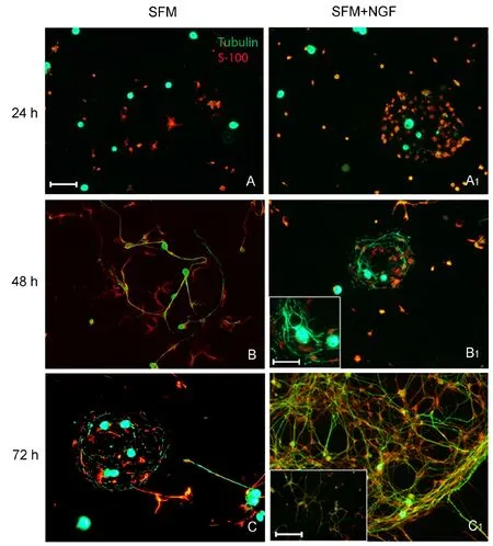
Figure 1 Dorsal root ganglia-derived dissociated cells spontaneously aggregate in vitro forming cell clusters.
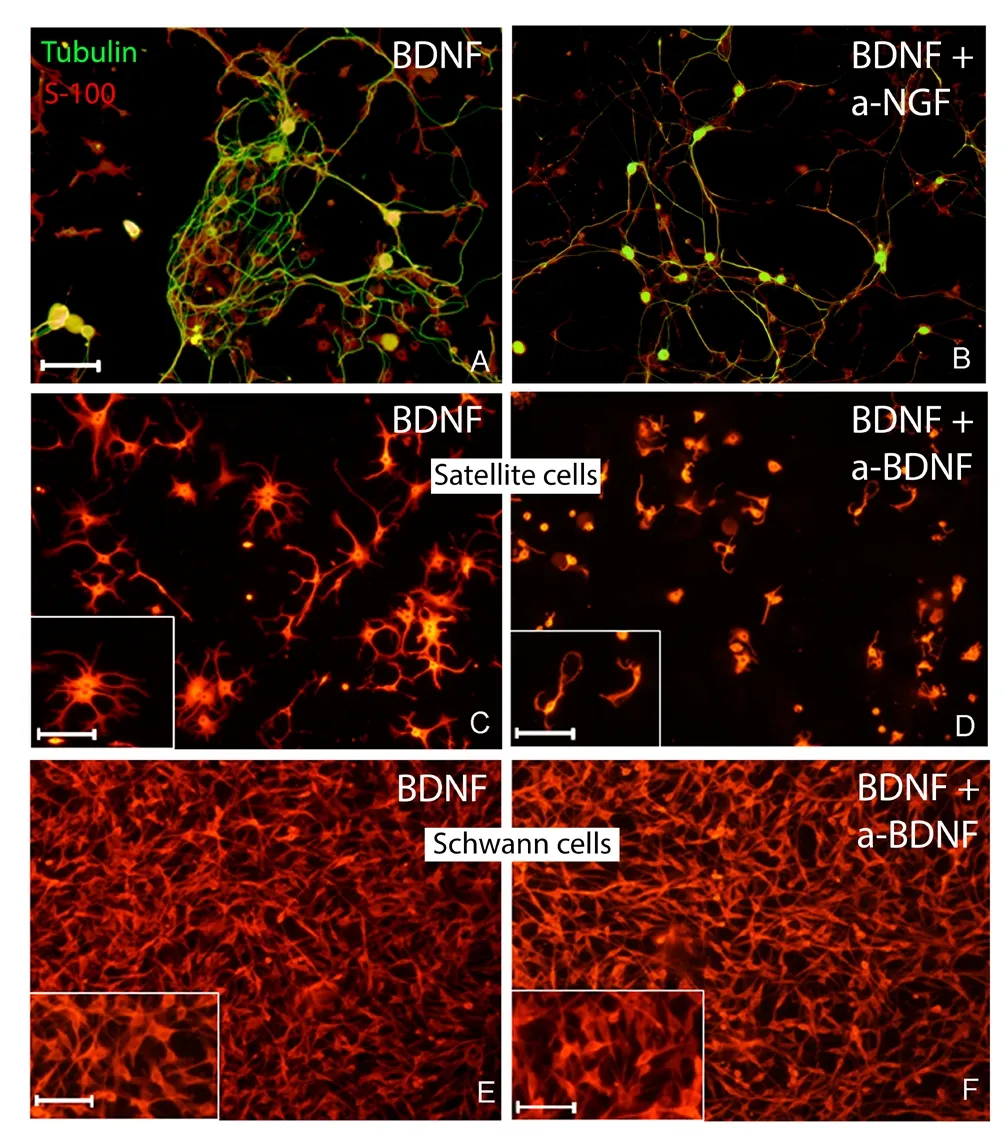
Figure 2 BDNF enhances cell cluster formation via activation of the satellite cells.
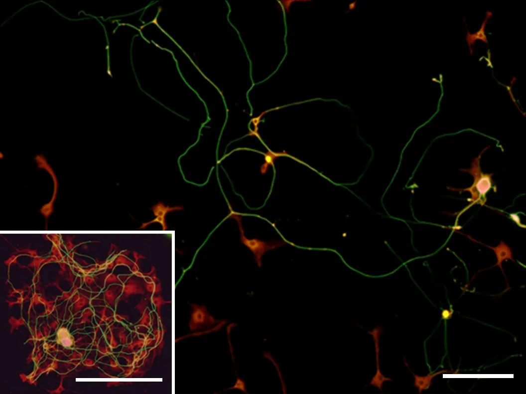
Figure 3 NT-3 enhances ordinate and oriented axonal elongation.
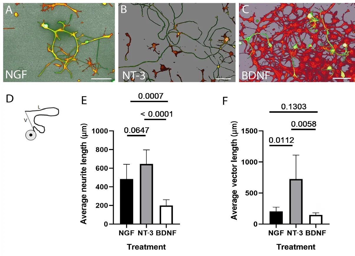
Figure 4 Quantification of neurite growth in dissociated cells.
The effect of BDNF on the neurite growth was similar to that of NGF. Dissociated cells were able to spontaneously aggregate and the neurite growth on both DRG models showed a lack of linearity. The 24-hour delay in the cluster formation together with the ability of anti-NGF antibody to null the effect of BDNF may suggest an indirect effect of BDNF on the growth of sensory fibers. Although we know that BDNF may have a preferential motor profile as previously suggested by Santos and colleagues (2016), we speculate that BDNF may enhance the release of NGF in the milieu via activation of the satellite glial cells (SGC). BDNF acting upstream to NGF would explain the delayed formation of cell cluster and the tortuosity of fiber growth. Little is known about SGC physiology and their interactions with neurons. Quantitative studies on several species showed that the number of SGC per neuron increases in proportion to the neuron’s volume (Pannese, 1981; Ledda et al., 2004; Haberberger et al., 2019), which is consistent with the idea that SGC support the neurons metabolically.
The re-clustering behavior of ganglionic cells in vitro has been mentioned before (Pannese and Procacci, 2002; Whitlon et al., 2009) and the consensual conclusion of these descriptive studies was that the formation of these clusters can be reconducted to the cross-talking between neurons and glia. Interestingly, the cross-talking between the sensory neurons and its supportive SGC modulates the morphology of both cells phenotypes, thus enhancing the outgrowth of slender projections from the nerve cell body surface and promoting neurite growth (Pannese and Procacci, 2002; Whitlon et al., 2009; Retamal et al., 2017). Like other glial cells, such as Schwann cells and astrocytes, SGC have numerous receptors for neurotransmitters and other bioactive molecules (Hanani, 2005; Jabs et al., 2005; Del Rio et al., 2015; Retamal, et al., 2017). Thus, it is possible that the cross-talk between sensory ganglia (neurons-neurons, neurons-SGC, and SGC-SGC) serves as a site for the modification of neuronal signals in the sensory pathway (Takeda et al., 2009; Huang et al., 2013).
Satellite cells express a high level of p75 receptor (Pannese, 1981; Dublin and Hanani, 2007; Koike et al., 2019) and TrkA (Pannese and Procacci 2002; Neto et al., 2017; Haberberger et al., 2019; Koike et al., 2019). Therefore, if BDNF enhances the metabolism of satellite cells and these cells express a high level of p75 as well as the NGF-specif ic receptor TrkA, then the satellite cells may be able to internalize NGF intracellularly as previously suggested by Pannese and Procacci (2002). The satellite cell itself or cross-talking with neurons (Deng et al., 2000; Chan et al., 2004; Huang et al., 2013; Del Rio et al., 2015; Retamal et al., 2017) may then release NGF in the milieu creating the environment that favors cell re-clustering formation and tortuous axonal growth. Ultimately, the neuron-supporting activity of the satellite cells may impede sensory axons to grow and extend outside of the ganglia.
Even though satellite cells and Schwann cells share many characteristics and both function as attendant cells to the neurons (Pannese et al., 1994; Huang et al., 2013), Schwann cells do not seem to be affected by BDNF. The expression of neurotrophin receptors is different between the two glial cell populations. Schwann cells in contact with axonal processes express very few if any p75 receptors. However, p75 is highly expressed in the absence of axons (Taniuchi et al., 1986; DiStefano and Johnson, 1988; Yamamoto et al., 1993; Tomita et al., 2007; Perez et al., 2019). It seems fair to speculate that in the case of peripheral nerve regeneration, Schwann cells which act as a conduit for axon regrowth, start expressing higher levels of p75 to bind and internalize NGF which would act as the target chemo-attractant source to guide and enhance the axonal growth. To our knowledge, no one thus far has reported the expression of TrkA receptors in Schwann cells of adult mammals (Althaus and Richter-Landsberg, 2000) which may affect the ability to internalize NGF and explain the different behaviors of these cells in the presence of BDNF.
Finally, out of the three members of the neurotrophin family considered in this study, our data identif ied NT-3 as the growth factor that plays a role in a linear and oriented axonal elongation. Both the dissociated ganglionic cells and the DRG explant models showed that NT-3 and NGF promoted axonal growth, however, NT-3 only confers linear orientation to growing neurites.
Conclusions
In agreement with Allodi et al., (2012), our data suggest a more prosensory role of NT-3 and NGF compared to BDNF. The most novel and clinically relevant aspect of our data is the effect of each factor on the linearity of growth. While NT-3 allows for a more linear elongation of growing neurites, NGF results in convoluted bundles of neurites winding around the neuronal cell body with less direction of growth. BDNF does not seem to have a direct effect on the linearity of sensory fiber growth. This finding not only adds some more to the level of understanding on the biochemistry of different growth factor members but also provides evidence to support possible applications in the field of regenerative medicine.
Acknowledgments:We sincerely thank Dr. Maria Giuseppina Robecchi for her help and support in experimental procedures, data analysis, and proof-reading the paper. We thank Ms. Kathryn Eggert and Mrs. Jennifer Lee for proof-reading the paper. We also thank the imaging core facility at Midwestern University for the technical support.
Author contributions:Study design: MF, IP; project supervisor and study coordinator: MF, SG, IP; experimental model establishment: MF, AG, AF, LM; experiment implementation: AG, LM, AF, GN; data collection and analysis: MF, AG, LM, AF, GN; result interpretation: MF, SG, IP; paper writing: MF. All authors approved the final version of this paper.
Conf licts of interest:The authors declare that they have no conf licts of interest.
Financial support:This study was supported by the research start-up and the MWU’s intramural grant (to MF) and the Italian MURST-MIUR foundation (to SG and IP).
Institutional review board statement:Ethical approval was obtained from the Regione Piemonte Animal Ethics Committee ASLTO1 (file # 864/2016-PR) on September 14, 2016.
Copyright license agreement:The Copyright License Agreement has been signed by all authors before publication.
Data sharing statement:Datasets analyzed during the current study are available from the corresponding author on reasonable request.
Plagiarism check:Checked twice by iThenticate.
Peer review:Externally peer reviewed.
Open access statement:This is an open access journal, and articles are distributed under the terms of the Creative Commons Attribution-Non-Commercial-ShareAlike 4.0 License, which allows others to remix, tweak, and build upon the work non-commercially, as long as appropriate credit is given and the new creations are licensed under the identical terms.

- 中國神經(jīng)再生研究(英文版)的其它文章
- Recovery of an injured ascending reticular activating system with recovery from a minimally conscious state to normal consciousness in a stroke patient: a diffusion tensor tractography study
- Resveratrol corrects aberrant splicing of RYR1 pre-mRNA and Ca2+ signal in myotonic dystrophy type 1 myotubes
- Interleukin-18 levels in the hippocampus and behavior of adult rat offspring exposed to prenatal restraint stress during early and late pregnancy
- EndoN treatment allows neuroblasts to leave the rostral migratory stream and migrate towards a lesion within the prefrontal cortex of rats
- A double-network hydrogel for the dynamic compression of the lumbar nerve root
- The Akt/glycogen synthase kinase-3β pathway participates in the neuroprotective effect of interleukin-4 against cerebral ischemia/reperfusion injury