
Inverted Y ureteral duplication with an ectopic ureter and multiple urinary calculi:A case report
2022-03-15 07:12:18WenXinYeLiGangRenLiChen
World Journal of Clinical Cases 2022年4期
INTRODUCTION
Ureteral duplication is defined as two ureters in the ipsilateral urinary system.A ureter with an abnormal positioning is called an ectopic ureter,and it often has an orifice outside the bladder triangle in duplicated ureteral malformations.Inverted Y duplicated ureteral malformation with ectopic ureteral calculi is uncommon in the literature,and only a few cases have been reported[1].There are no relevant reports on cases with multiple urinary calculi in the renal pelvis and ureters.
CASE PRESENTATION
Chief complaints
A 36-year-old man was admitted to the hospital with complaints of right lumbar pain for one month.
When the bell rings get up quickly and eat your breakfast, and you will find the same horse waiting to take you home; but remember that you must never expect to see my palace again
History of present illness
The patient saw a doctor due to right lumbar pain for one month.He had no gross hematuria,chills,fever,or urinary incontinence.No history of trauma was reported.
History of past illness
The patient had no notable previous medical history.
Personal and family history
Dummling went and cut down the tree, and when it fell there was a goose19 sitting in the roots with feathers of pure gold.20 He lifted her up, and taking her with him, went to an inn where he thought he would stay the night. Now the host had three daughters,.21 who saw the goose and were curious to know what such a wonderful bird might be, and would have liked to have one of its golden feathers.
Physical examination
I began to understand what he meant one evening when my mother gave me money to phone a message to my father at the small upholstery shop where he had found temporary work. I went to a pay phone and dialed the number.
The patient was conscious.His vital signs were as follows:body temperature,36.5 °C;pulse,82/min;respiration rate,18/min;blood pressure,130/72 mmHg;right kidney area percussion pain was positive;and abdominal findings were unremarkable.
Laboratory examinations
Excluding the operative contraindications,a right transurethral ureteroscopic holmium laser lithotripsy was performed on November 10,2020.No ectopic ureteral opening was found in the urethra or bladder by endoscopy,and ureteroscopy was used to enter the right ureter through a normal bladder opening along the guidewire.There was a stone in the upper ureter,approximately 1.5 cm × 1.0 cm in size,and a stone was present in the lower calyx of the right kidney,approximately 1.0 cm × 1.0 cm in size.A holmium laser was used to pulverize the stones.The ureteral sheath was placed into the right ureter,and the contrast agent was injected.The ectopic ureter was observed by fluoroscopy using a C model arm X-ray machine.A duplicated ureteral bifurcation could be observed with a flexible ureteroscope (Figure 4),and the intraoperative diagnosis was an inverted Y ureteral duplication and ectopic ureteral multiple stones.We informed the patient’s family members regarding the intraoperative findings and recommended that he undergo laparoscopic ectopic ureterectomy.Unfortunately,the family temporarily refused laparoscopic surgery.
The patient denied any family history and had no notable history.
Inverted Y ureteral duplication is a rare congenital malformation of the ureter.There are a few reports in the literature.This malformation occurs when two distal ureters fuse into one and then enter the renal pelvis.Similar to other types of duplicated ureteral malformations,the incidence of inverted Y ureteral duplication in women is higher than that in men[2].In inverted Y ureteral duplication,two distal ureters can open into the bladder.Usually,one ureter opens into the bladder,and the other ureter has an ectopic opening or the end of the ureter has atresia.The mechanism of its formation is not clear,but it is currently thought that its formation is related to the fusion of two independent ureteral bud tips into one tube before connecting to the metanephros[3].
Imaging examinations
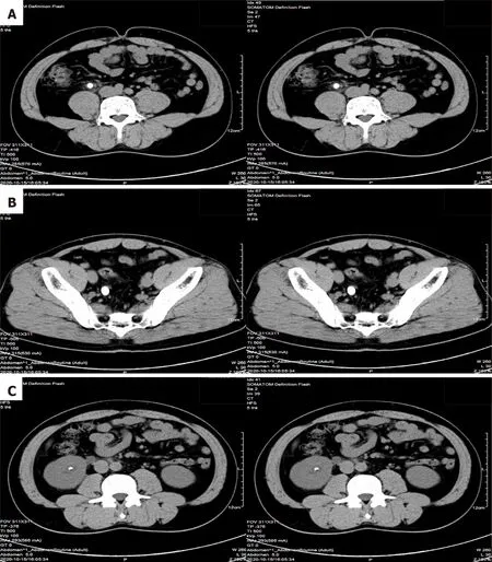
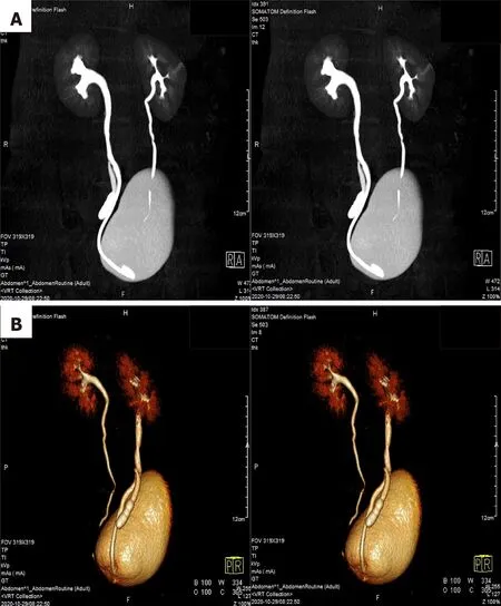
Computed tomography scans revealed one stone in the right upper ureter,multiple stones in the right lower ureter,and other stones in the right kidney (Figure 1).Computed tomography urography and three-dimensional reconstructions revealed that the middle of the right ureter was divided into two branches,one ureter descended to the bladder with a normal bladder opening and the other ureter descended to the vicinity of the right seminal vesicle with an ectopic opening,suggesting duplicated ureteral malformation with an ectopic ureter (Figure 2).
FINAL DIAGNOSIS
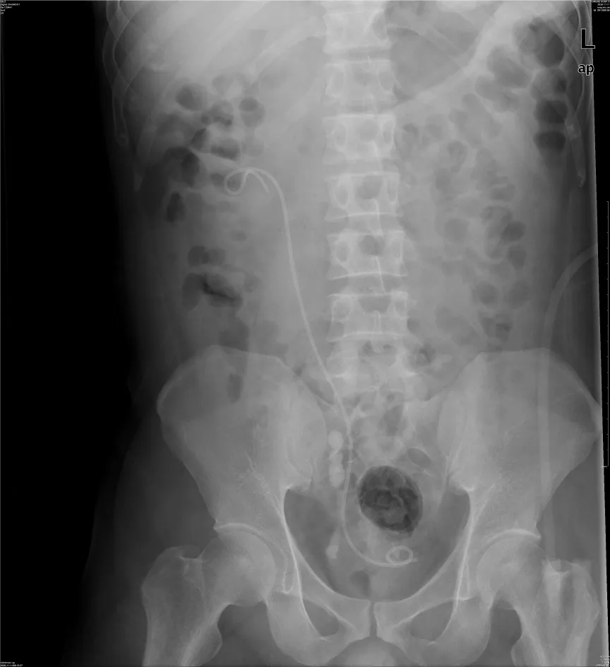
The KUB was rechecked on the second day after surgery (Figure 5),and then the patient was discharged.The double J tube was removed 3 wk after the operation.The patient did not feel any discomfort after one year of follow-up.
TREATMENT
Laboratory data were unremarkable except for urine red blood cells (+++) on urinalysis.
OUTCOME AND FOLLOW-UP
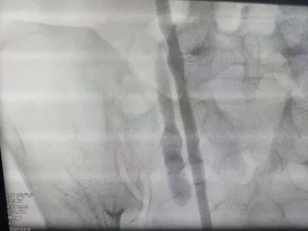
From the imaging findings and intraoperative retrograde ureterography,the final diagnosis was as follows:A right inverted Y duplicated ureteral malformation;right fusion broncho-ureteral stones;multiple stones in the ectopic ureter;and a right kidney stone (Figure 3).
DISCUSSION
18. Ordered one of them to go and examine the house: The robbers do not work together like the animals, and the captain has no reservations about exposing a member of his band to danger (Tatar 156). The animals, on the hand, work to protect each other, not only from the robbers but from the owners.Return to place in story.
The clinical symptoms of patients with an inverted Y duplicated ureteral malformation are diverse,and they mainly depend on the location of the ectopic ureteral opening.The opening of ectopic ureters into the vulva or reproductive organs can cause incontinence[4,5];the opening of ectopic ureters into the bladder and form ureteral cysts can cause bladder outlet obstruction[6];the opening of ectopic ureters into the seminal vesicles can cause discomfort in the lower abdomen or infertility[7];and ureteral stones in the ectopic ureter may cause abdominal pain or gross hematuria[1].
An inverted Y duplicated ureteral malformation cannot be diagnosed by clinical symptoms and is mainly diagnosed by ureterography.Retrograde ureterography routinely develops by X-rays and can also develop by contrast-enhanced ultrasound[8].
There is no standard for the treatment of inverted Y duplicated ureteral malformations or their comorbidities.Asymptomatic patients with inverted Y duplicated ureteral malformations can be followed up for observation.For comorbidities caused by ectopic ureters,corresponding treatment can be performed.When ectopic ureteral cysts cause bladder outflow obstruction,removal of the endoscopic cyst is feasible[9].For ectopic ureters that cause incontinence or intraureteral stones,laparoscopic ectopic ureterectomy is feasible[10],and replantation of the ureter can also be considered[1].
Everything in the right place! the pedlar said when he had atlast safely got out of Sodom and Gomorrah, as he called it. Theopen high road is my right place; up there I did not feel at ease. The little maid, who was still watching the geese, nodded kindlyto him as he passed through the gate.
In this case,the inverted Y ureteral duplication combined with multiple urinary calculi not only contributed to the stones in the renal pelvis and the fused bronchoureter,but also contributed to the development of multiple stones in the ectopic ureter.It is not clear whether the ectopic ureteral stones developed from the kidney or another primary site.Fused broncho-ureteral stones can cause hydronephrosis,which can manifest as lumbar pain,while ectopic ureteral stones may not cause clinical symptoms and are often ignored.The methods for treating urinary calculi in different parts of the urinary tract are different.In this case,a transurethral ureteroscopic holmium laser lithotripsy was used to treat fused broncho-ureteral calculi and intrarenal pelvic calculi,and the results were satisfactory.Our only regret is that the patient refused to treat the ectopic ureteral stones,and surgical specimens could not be obtained.During the one-year follow-up,the patient had no obvious symptoms,such as lumbar pain.However,because of long-term entrapment and stimulation by urinary calculi,urinary calculi can cause urinary tract infections and can increase the risk of cancer,and it is recommended that patients with an ectopic ureter have it removed as soon as possible during follow-up.The most minimally invasive and effective method currently reported in the literature is laparoscopic ectopic ureterectomy.
CONCLUSION
For patients with an inverted Y ureteral duplication and multiple urinary calculi,it is important to clarify the type of ureteral malformation and the location of the stones,and these identifications can have a positive effect on the treatment.For stones in the renal pelvis and ureters with normal openings,endoscopic lithotripsy can be used to achieve good results.For stones in an ectopic ureter,it is difficult to find or locate the ectopic ureteral opening,and most patients are advised to have the ectopic ureter removed.At present,laparoscopic ectopic ureterectomy is a common procedure,and the consequent outcome is usually good.
 World Journal of Clinical Cases2022年4期
- World Journal of Clinical Cases的其它文章
- Surgical treatment of acute cholecystitis in patients with confirmed COVID-19:Ten case reports and review of literature
- Rituximab as a treatment for human immunodeficiency virusassociated nemaline myopathy:What does the literature have to tell us?
- Eustachian tube involvement in a patient with relapsing polychondritis detected by magnetic resonance imaging:A case report
- Endoscopic clipping for the secondary prophylaxis of bleeding gastric varices in a patient with cirrhosis:A case report
- Inflammatory myofibroblastic tumor after breast prosthesis:A case report and literature review
- Langerhans cell histiocytosis presenting as an isolated brain tumour:A case report