
Quantitative volumetric analysis of the optic radiation in the normal human brain using diffusion tensor magnetic resonance imaging-based tractography
2014-03-24 02:51:18DongHoonLeeJiWonParkCheolPyoHong
中國(guó)神經(jīng)再生研究(英文版) 2014年3期
Dong-Hoon Lee, Ji-Won Park, Cheol-Pyo Hong
1 Center for Medical Metrology, Division of Convergence Technology, Korea Research Institute of Standards and Science (KRISS), Daejeon, Republic of Korea
2 Department of Radiological Science, College of Health Science, Yonsei University, Wonju, Republic of Korea
3 Department of Physical Therapy, College of Medical Science, Catholic University of Daegu, Daegu, Republic of Korea
Quantitative volumetric analysis of the optic radiation in the normal human brain using diffusion tensor magnetic resonance imaging-based tractography
Dong-Hoon Lee1,2, Ji-Won Park3, Cheol-Pyo Hong1
1 Center for Medical Metrology, Division of Convergence Technology, Korea Research Institute of Standards and Science (KRISS), Daejeon, Republic of Korea
2 Department of Radiological Science, College of Health Science, Yonsei University, Wonju, Republic of Korea
3 Department of Physical Therapy, College of Medical Science, Catholic University of Daegu, Daegu, Republic of Korea
To attain the volumetric information of the optic radiation in normal human brains, we performed diffusion tensor imaging examination in 13 healthy volunteers. Simultaneously, we used a brain normalization method to reduce individual brain variation and increase the accuracy of volumetric information analysis. In addition, tractography-based group mapping method was also used to investigate the probability and distribution of the optic radiation pathways. Our results showed that the measured optic radiation fi ber tract volume was a range of about 0.16% and that the fractional anisotropy value was about 0.53. Moreover, the optic radiation probability fi ber pathway that was determined with diffusion tensor tractography-based group mapping was able to detect the location relatively accurately. We believe that our methods and results are helpful in the study of optic radiation fi ber tract information.
nerve regeneration; optic radiation; diffusion tensor imaging; diffusion tensor tractography; magnetic resonance imaging; volumetric analysis; probability map; group mapping; visualization; individual variation; neural regeneration
Lee DH, Park JW, Hong CP. Quantitative volumetric analysis of the optic radiation in the normal human brain using diffusion tensor magnetic resonance imaging-based tractography. Neural Regen Res. 2014;9(3):280-284.
Introduction
The human visual system consists of two major components, sensory input organs, such as the retina in the eyes, and the visual pathway[1]. The object information that is captured through the eyes is transmitted by the retina and their axons, which are comprised of nerve fi bers, to the optic chiasm[1-2]. The visual pathway continues to the lateral geniculate nucleus[1-4]. The optic radiation is a dense fi ber tract that emerges from the lateral geniculate nucleus and continues to the occipital visual cortex[1-4]. Especially, the optic radiation is an important fi ber structure that conveys visual information from the lateral geniculate nucleus to the primary visual cortex in the occipital lobe. In recent years, many studies have introduced the anatomical location and features of the optic radiation[1-10]. Knowing such above-mentioned characteristic information of the optic radiation before operation is important for surgery of the temporal lobe, longitudinal study of patients with optic neuritis, and evaluation of visual function in preterm infants[5,7-8,10-12]. Most of these studies have used diffusion tensor imaging, which is a magnetic resonance imaging technique that is used to examine the directional properties of the diffusion of water molecules[13-20]. In particular, several studies have been published on these topics related to the optic radiation, and these have described the characteristic information obtained from diffusion tensor tractography, which is derived from diffusion tensor imaging and is a robust technique that is used for visualizing and evaluating white matter fiber direction in the human brain[1-2,7-12,21]. However, these studies have focused on the anatomical characteristics of optic radiation fi ber tracts in individual brains and on comparisons of the anatomical characteristics of the optic radiation fi ber tracts between patient and control groups. Therefore, to the best of our knowledge, no diffusion tensor tractography studies of the volumetric information of optic radiation have been conducted without individual brain structure variation.
In the current study, we attempted to analyze the volumetric information of the optic radiation and to investigate the characteristics of the optic radiation in normal human brain with diffusion tensor tractography.
Results
Quantitative data
The values of the counted voxel numbers for the normalized optic radiation fi ber tracts, and the percentage of the counted voxel numbers that were divided by whole voxel numbers in the Montreal Neurologic Institute (MNI) echo-planar imaging (EPI) template are shown in Table 1. These values represent the ratio of the optic radiation fi ber tract volumetricinformation in both hemispheres of normal human brain.
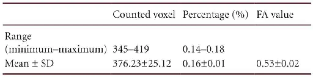
Table 1 The counted voxel numbers that were part of the extracted optic radiation fiber tracts, the calculated percentage, and the fractional anisotropy (FA) value for each subject
Probability map
The optic radiation probability pathway map, which represents the degree of overlapping optic radiation fi ber tracts of all subjects, was overlaid onto a standard MNI EPI template (Figure 1).
Discussion
In the current study, we analyzed the volumetric information of optic radiation fi ber tracts in the human brain with brain normalization methods with a standard brain template image. In addition, we visualized a probability fi ber pathway map in order to investigate the probability and distribution of the optic radiation pathway. Since the introduction of diffusion tensor imaging and fi ber tracking methods, these techniques have been widely used to elucidate the anatomical structure and to perform quantitative analyses of neural fi ber tracts at the subcortical level[16-17,19,22-38]. Many studies have been conducted on the optic radiation, although the majority of these studies have focused on examining the anatomical structures or landmarks of surgical planning in patients[3,4,9,11-12,21,25]. Schoth et al.[21]have reported anisotropy changes in blind humans compared to healthy control subjects with diffusion tensor tractography. Winston et al.[4]have described the use of optic radiation fi ber tractography in epilepsy surgical planning and anterior temporal lobe resections. Ciccarelli et al.[7]have indicated anatomical changes after optic neuritis, and they showed that optic radiation fiber tract location was different between patient and normal control groups. Moreover, some quantitative fi ber tracking analyses and approaches have been used in patients with arteriovenous malformations and premature newborns in order to evaluate the optic radiation fi ber tracts[5-6,26]. In the quantitative analyses, FA values, diffusivity indices, and leftto-right asymmetry indices were measured in these patients.
In this study, we measured the volumetric information of the optic radiation fi ber tracts in order to determine the proportion of optic radiation fi ber tracts in the whole brain of the normal human brain. The extracted optic radiation fiber tracts were normalized to a standard brain template, the MNI EPI template, in order to remove the individual differences of all of the subjects and to allow for a direct comparison of our volumetric analysis results under the same conditions. The results of this study demonstrated that the optic radiation fi ber tract had a tiny volume of information that was less than 1% of visual performance compared to the volume information of the whole brain. Moreover, the optic radiation probability fi ber pathway that was determined with diffusion tensor tractography-based group mapping was able to detect location with relative accuracy.
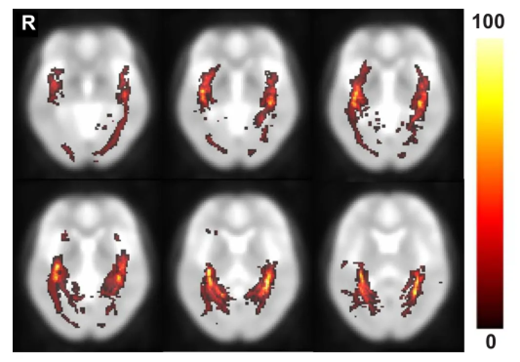
Figure 1 The results of the optic radiation probability pathway map in healthy subjects.
This study had some limitations. First, a limited number of subjects were enrolled in this study. Based on our results, the 13 healthy subjects exhibited a similar tendency for the percentage of counted voxel numbers. However, these fi ndings are dif fi cult to generalize to our results. In a future study, we plan to study a larger number of subjects. Second, the setting of the region of interest is a user-dependent process. This problem can be solved through fi ber tracking that is combined with functional magnetic resonance imaging examinations of visual stimulation in a future study. Third, we evaluated only the optic radiation fi ber tract of all of the optic fibers in the human brain. Moreover, the Fuzzy art with Add Clustering Technique (FACT) algorithm, which was used in this study, is used for limited fi ber tracking and reconstruction. The FACT algorithm was considered for determining the dominant fiber direction, and the largest eigenvalue components in each voxel were used as an indicator of fi ber orientation. Therefore, a fi ber-crossing region or a voxel that was eligible for various fi ber directions might have resulted in some erroneous points in the FACT algorithm-based fiber tracking. Further study is needed with other fi ber tracking and reconstruction algorithms, such as probabilistic approaches or with an advanced fast marching algorithm. The probabilistic fi ber tracking algorithm allows for the direction of the tensor in the voxel to be multidirectional, but it is not limited to a dominant direction, as is the FACT algorithm. The advanced fast marching takes into account all of the information that is contained in the diffusion tensor[2]. Thereby, every tensor is classi fi ed as linear, planar, or spherical ellipsoid[2,27]. In other words, the method that was described above is able to evaluate brain areas thatcontain fiber-crossing regions, such as the optic chiasm. They have important implications for more accurate fiber tracking for all of the optic nerve that is not limited to the optic radiation, which is a topic for future research.
In conclusion, we analyzed and measured the volume information of the optic radiation fi ber tract in the normal human brain and found that the optic radiation fi ber tract had a relatively small range of volume information compared to that of the whole brain. To the best of our knowledge, this is the first volumetric analysis study of the optic radiation fi ber tract with diffusion tensor tractography and of wholebrain volume information. We believe that our approaches provide preliminary data for researchers who are studying treatments for patients with diseases that are related to the visual pathway in the brain.
Subjects and Methods
Design
An observational neuroimaging study.
Time and setting
This experiment was performed at the Department of Physical Medicine and Rehabilitation, Yeungnam University Hospital, Republic of Korea in June 2009.
Subjects
Thirteen healthy subjects (men, 6; age, 35 ± 2.16 years) were recruited into this study through advertisements. They had no previous history of neurological disease, optic nerve pathology, head injury, or physical disease. All subjects understood the purpose of the study and provided written informed consents prior to their participation. This study was approved by the Institutional Review Board of Yeungnam University Hospital in Republic of Korea.
Methods
Diffusion tensor imaging acquisition
All diffusion tensor imaging datasets were acquired with a 1.5-T magnetic resonance imaging system (Gyroscan Intera, Philips Healthcare, Best, the Netherlands) with a six-channel phased-array sensitivity-encoding (SENSE) head coil by using single-shot echo-planar imaging (EPI) with parallel acquisition in the transverse plane. The imaging parameters were as follows: repetition time/echo time, 10,726/76 ms; matrix, 128 × 128; fi eld of view, 221 mm; slice thickness, 2.3 mm; and reduction factor for SENSE, 2. Diffusion weighting was applied along 32 non-collinear diffusion-sensitizing gradients with a b-value of 1,000 s/mm2. We obtained 63–67 contiguous transverse slices that covered the entire brain with no slice gaps. During the acquisition, radiologists routinely checked for gross head movement or other motion in real time[25].
Diffusion tensor imaging analysis
Eddy current corrections of the diffusion tensor imaging data were performed with the tool in FSL 4.0.1 (Analysis Group, FMRIB, Oxford, UK) with a 12-parameter affine registration[4-5,12,25,39]. Each diffusion-weighted image was registered to non-diffusion weighted images (b = 0). The diffusion tensor imaging datasets were analyzed with DtiStudio 3.0.3 (Department of Radiology, Johns Hopkins University, Baltimore, MD, USA), which was based on the fi ber assignment by the continuous tracking (FACT) algorithm[22,40-43]. Propagation in each fi ber tract was terminated if a voxel with a fractional anisotropy (FA) value less than 0.25 was reached or if the turning angles of 2 consecutive vectors were over70° during tracking[10]. An FA threshold of 0.25–0.35 for fiber tract reconstruction has been recommended by Mori et al. and Stieltjes et al.[10,23,44]. A relatively large angle threshold was used so that the optic radiation course in acute angles could be examined[10,26]. The region of interest was manually drawn in both lateral geniculate nucleus on the transverse plane at the level of the transition from the posterior limb of the internal capsule to the cerebral peduncle on a color-coded FA map, and the volume of the region of interest was standardized for all subjects (9 voxels)[3,7]. The color-coded FA map showed the fi ber pathway directions with 3 colors as follows: red (left-right direction), green (anterior-posterior), and blue (superior-inferior)[45-46].
Quantitative measurements and probability map of the optic radiation
After both optic radiation fiber tracts were extracted with the region of interest described above, we performed quantitative measurements and created a probability pathway map of the optic radiation. These methods were conducted based on the brain normalization method in order to avoid individual brain variation. The procedure is shown in Figure 2 and is described as follows. (1) Each subject’s non-diffusion image (b = 0 image) was co-registered to the standard Montreal Neurologic Institute (MNI) space with an EPI template with SPM2 (Wellcome Department of Cognitive Neurology, London, UK). (2) The transformation matrices for each subject that were created with the coregistration process in (1) were then applied to the extracted optic radiation fi ber tracts of each subject for normalization to the MNI space. (3) For the quantitative measurements, the voxels that the optic radiation fi ber tract passed through in the normalized brain of each subject were counted with ImageJ (US National Institutes of Health, Bethesda, MD, USA) software. We calculated the percentage of the optic radiation fi ber tracts with the counted voxel numbers divided by the whole-brain voxel numbers in the MNI EPI template. (4) In order to assess optic radiation fi ber tracts quantitatively, the FA values in the extracted optic radiation fi ber tracts for each subject were measured. (5) Finally, each individual normalized optic radiation was averaged pixel-by-pixel and overlaid on the MNI EPI template in order to obtain and investigate the optic radiation probability pathway map with MRIcro (Chris Rorden, USA, http://www.mricro.com) software.
Author contributions:Lee DH participated in study conception, design and analysis and manuscript development. Park JW contributed to data acquisition and analysis. Hong CP contributed tostudy design, manuscript development, oversight and research supervision. All authors approved the final version of this paper.
Con fl icts of interest:None declared.
Peer review:This study was designed to provide information about the average size and degree of diffusion anisotropy of the optic radiation in addition to a probability map describing the likelihood of optic radiation pathway. The proposed analysis was applied to 13 healthy subjects with a mean age of 35 years and the results were demonstrated according to the analysis. This type of analysis and results are considered to be an addition to the field of diffusion tensor imaging because of inaccurate diffusion tensor imaging measurement, approximate tensor model and relatively low resolution diffusion tensor imaging. The difficulties are to verify the segmented optic radiation using various tractography segmentation algorithms. Quantitative measurements of optic radiation probability map and the estimated optic radiation size and anisotropy can help identify healthy subjects and be used as a reference for comparing healthy to pathological subjects.
[1] El-Rafei A, Engelhorn T, W?rntges S, et al. A framework for voxel-based morphometric analysis of the optic radiation using diffusion tensor imaging in glaucoma. Magn Reson Imaging. 2011;29: 1076-1087.
[2] Staemp fl i P, Rienmueller A, Reischauer C, et al. Reconstruction of the human visual system based on diffusion tensor imaging fi ber tracking. J Magn Reson Imaging. 2007;26:886-893.
[3] Sherbondy AJ, Dougherty RF, Napel S, et al. Identifying the human optic radiation using diffusion imaging and fi ber tractography. J Vis. 2008;8:12.1-1211.
[4] Winston GP, Mancini L, Stretton J, et al. Diffusion tensor imaging tractography of the optic radiation for epilepsy surgical planning: a comparison of two methods. Epilepsy Res. 2011;97:124-132.
[5] Bassi L, Ricci D, Volzone A, et al. Probabilistic diffusion tractography of the optic radiations and visual function in preterm infants at term equivalent age. Brain. 2008;131:573-582.
[6] Berman JI, Glass HC, Miller SP, et al. Quantitative fi ber tracking analysis of the optic radiation correlated with visual performance in premature newborns. Am J Neuroradiol. 2009;30:120-124.
[7] Ciccarelli O, Toosy AT, Hickman SJ, et al. Optic radiation changes after optic neuritis detected by tractography-based group mapping. Hum Brain Mapp. 2005;25:308-316.
[8] Kolbe S, Bajraszewski C, Chapman C, et al. Egan, Diffusion tensor imaging of the optic radiations after optic neuritis. Hum Brain Mapp. 2012;33:2047-2061.
[9] Nilsson D, Starck G, Ljungberg M, et al. Intersubject variability in the anterior extent of the optic radiation assessed by tractography. Epilepsy Res. 2007;77:11-16.
[10] Yamamoto A, Miki Y, Urayama S, et al. Diffusion tensor fiber tractography of the optic radiation: analysis with 6-, 12-, 40-, and 81-directional motion-probing gradients, a preliminary study. Am J Neuroradiol. 2007;28:92-96.
[11] Powell HW, Parker GJ, Alexander DC, et al. MR tractography predicts visual fi eld defects following temporal lobe resection. Neurology. 2005;65:596-599.
[12] Winston GP, Daga P, Stretton J, et al. Optic radiation tractography and vision in anterior temporal lobe resection. Ann Neurol. 2012;71:334-341.
[13] Basser PJ, Mattiello J, LeBihan D. Estimation of the effective self-diffusion tensor from the NMR spin echo. J Magn Reson B. 1994;103:247-254.
[14] Basser PJ, Mattiello J, LeBihan D. MR diffusion tensor spectroscopy and imaging. Biophys J. 1994;66:259-267.
[15] Beaulieu C. The basis of anisotropic water diffusion in the nervous system-a technical review. NMR Biomed. 2002;15:435-455.
[16] Mori S, Crain BJ, Chacko VP, et al. Three-dimensional tracking of axonal projections in the brain by magnetic resonance imaging. Ann Neurol. 1999;45:265-269.
[17] Mori S, van Zijl PC. Fiber tracking: principles and strategies-a technical review. NMR Biomed. 2002;15:468-480.
[18] Pierpaoli C, Basser PJ. Toward a quantitative assessment of diffusion anisotropy. Magn Reson Med. 1996;36:893-906.
[19] Pierpaoli C, Jezzard P, Basser PJ, et al. Diffusion tensor MR imaging of the human brain. Radiology. 1996;201:637-648.
[20] Wakana S, Jiang H, Nagae-Poetscher LM, et al. Fiber tract-based atlas of human white matter anatomy. Radiology. 2004;230:77-87.
[21] Schoth F, Burgel U, Dorsch R, et al. Diffusion tensor imaging in acquired blind humans. Neurosci Lett. 2006;398:178-182.
[22] Jiang H, van Zijl PC, Kim J, et al. DtiStudio: resource program for diffusion tensor computation and fi ber bundle tracking. Comput Methods Programs Biomed. 2006;81:106-116.
[23] Mori S, Kaufmann WE, Davatzikos C, et al. Imaging cortical association tracts in the human brain using diffusion-tensor-based axonal tracking. Magn Reson Med. 2002;47:215-223.
[24] White ML, Zhang Y. Three-tesla diffusion tensor imaging of Meyer’s loop by tractography, color-coded fractional anisotropy maps, and eigenvectors. Clin Imaging. 2010;34:413-417.
[25] Yogarajah M, Focke NK, Bonelli S, et al. Defining Meyer’s loop-temporal lobe resections, visual field deficits and diffusion tensor tractography. Brain. 2009;132:1656-1668.
[26] Okada T, Miki Y, Kikuta K, et al. Diffusion tensor fiber tractography for arteriovenous malformations: quantitative analyses to evaluate the corticospinal tract and optic radiation. Am J Neuroradiol. 2007;28:1107-1113.
[27] Westin CF, Maier SE, Mamata H, et al. Processing and visualization for diffusion tensor MRI. Med Image Anal. 2002;6:93-108.
[28] Seo JP, Lee MY, Kwon YH, et al. Delayed gait recovery in a stroke patient. Neural Regen Res. 2013;8:1514-1518.
[29] Li J, Chen X, Zhang J, et al. Intraoperative diffusion tensor imaging predicts the recovery of motor dysfunction after insular lesions. Neural Regen Res. 2013;8:1400-1409.
[30] Kuhnt D, Bauer MH, Sommer J, et al. Optic radiation fi ber tractography in glioma patients based on high angular resolution diffusion imaging with compressed sensing compared with diffusion tensor imaging-initial experience. PLoS One. 2013;8:e70973
[31] Surova Y, Szczepankiewicz F, Latt J, et al. Assessment of global and regional diffusion changes along white matter tracts in Parkinsonian disorders by MR tractography. PLoS One. 2013;8:e66022.
[32] Seo JP, Jang SH. Traumatic thalamic injury demonstrated by diffusion tensor tractography of the spinothalamic pathway. Brain Inj. 2013;27:749-753.
[33] Cauley KA, Filippi CG. Diffusion-tensor imaging of small nerve bundles: cranial nerves, peripheral nerves, distal spinal cord, and lumbar nerve roots- clinical applications. Am J Roentgenol. 2013;201:W326-335.
[34] Yeo SS, Jang SH. Corticospinal tract recovery in a patient with traumatic transtentorial herniation. Neural Regen Res. 2013;8:469-473.
[35] El-Rafei A, Engelhorn T, Warntges S, et al. Glaucoma classi fi cation based on visual pathway analysis using diffusion tensor imaging. Magn Reson Imaging. 2013;31:1081-1091.
[36] Seo JP, Choi BY, Chang CH, et al. Diffusion tensor imaging fi ndings of optic radiation in patients with putaminal hemorrhage. Eur Neurol. 2013;69:236-241.
[37] Wang S, Qiu D, So KF, et al. Radiation induced brain injury: assessment of white matter tracts in a pre-clinical animal model using diffusion tensor MR imaging. J Neurooncol. 2013;112:9-15.
[38] Yeo SS, Kim SH, Kim OL, et al. Optic radiation injury in a patient with traumatic brain injury. Brain Inj. 2012;26:891-895.
[39] Smith SM, Jenkinson M, Woolrich MW, et al. Advances in functional and structural MR image analysis and implementation as FSL. Neuroimage. 2004;23 Suppl 1:S208-219.
[40] Krishnan AP, Asher IM, Davis D, et al. Evidence that MR diffusion tensor imaging (tractography) predicts the natural history of regional progression in patients irradiated conformally for primary brain tumors. Int J Radiat Oncol Biol Phys. 2008;71:1553-1562.
[41] Kim CH, Koo BB, Chung CK, et al. Thalamic changes in temporal lobe epilepsy with and without hippocampal sclerosis: a diffusion tensor imaging study. Epilepsy Res. 2010;90:21-27.
[42] Lima M, Yamamoto A, Brion V, et al. Reduced-distortion diffusion MRI of the craniovertebral junction. Am J Neuroradiol. 2012;33:1321-1325.
[43] Wiltshire K, Concha L, Gee M, et al. Corpus callosum and cingulum tractography in Parkinson’s disease. Can J Neurol Sci. 2010; 37:595-600.
[44] Stieltjes B, Kaufmann WE van Zijl PC, et al. Diffusion tensor imaging and axonal tracking in the human brainstem. Neuroimage. 2001;14:723-735.
[45] Lee SK, Kim DI, Kim J, et al. Diffusion-tensor MR imaging and fiber tractography: a new method of describing aberrant fiber connections in development CNS anomalies. Radiographics. 2005;25:53-65.
[46] Huang H, Yamamoto Akria, Hossain MA, et al. Quantitative Cortical Mapping of Fractional Anisotropy in Developing Rat Brains. J Neurosci. 2008;28:1427-1433.
Copyedited by Ng WH, El-Rafei A, Li CH, Song LP, Liu WJ, Zhao M
10.4103/1673-5374.128223
Cheol-Pyo Hong, Ph.D., 267 Gajeong-Ro, Yuseong-Gu, Daejeon 305-340, Republic of Korea, dosagehong@kriss.re.kr.
http://www.nrronline.org/
Accepted: 2013-11-25
 中國(guó)神經(jīng)再生研究(英文版)2014年3期
- 中國(guó)神經(jīng)再生研究(英文版)的其它文章
- The Olig family affects central nervous system development and disease
- Short-term environmental enrichment exposure induces proliferation and maturation of doublecortin-positive cells in the prefrontal cortex
- Special function of nestin+neurons in the medial septum-diagonal band of Broca in adult rats
- Normalization of ventral tegmental area structure following acupuncture in a rat model of heroin relapse
- Acupuncture at Waiguan (SJ5) and sham points in fl uences activation of functional brain areas of ischemic stroke patients: a functional magnetic resonance imaging study
- Can amino-functionalized carbon nanotubes carry functional nerve growth factor?