
Optimal duration of percutaneous microballoon compression for treatment of trigeminal nerve injury
2014-03-24 02:10:11FuyongLiShuaiHanYiMaFuxinYiXinminXuYunhuiLiu
中國神經(jīng)再生研究(英文版) 2014年2期
Fuyong Li, Shuai Han, Yi Ma, Fuxin Yi, Xinmin Xu, Yunhui Liu
1 Department of Neurosurgery, Shengjing Hospital, China Medical University, Shenyang, Liaoning Province, China
2 Department of Neurosurgery, the First Hospital of China Medical University, Shenyang, Liaoning Province, China
3 Second Department of Neurosurgery, the People’s Hospital of Liaoning Province, Shenyang, Liaoning Province, China
4 First Department of Neurosurgery, the First Af fi liated Hospital, Liaoning Medical College, Jinzhou, Liaoning Province, China
Optimal duration of percutaneous microballoon compression for treatment of trigeminal nerve injury
Fuyong Li1, Shuai Han2, Yi Ma3, Fuxin Yi4, Xinmin Xu3, Yunhui Liu1
1 Department of Neurosurgery, Shengjing Hospital, China Medical University, Shenyang, Liaoning Province, China
2 Department of Neurosurgery, the First Hospital of China Medical University, Shenyang, Liaoning Province, China
3 Second Department of Neurosurgery, the People’s Hospital of Liaoning Province, Shenyang, Liaoning Province, China
4 First Department of Neurosurgery, the First Af fi liated Hospital, Liaoning Medical College, Jinzhou, Liaoning Province, China
Fuyong Li, Ph.D., now working at the Second Department of Neurosurgery, the People’s Hospital of Liaoning Province, China
Fuyong Li and Shuai Han contributed equally to this article.
Percutaneous microballoon compression of the trigeminal ganglion is a brand new operative technique for the treatment of trigeminal neuralgia. However, it is unclear how the procedure mediates pain relief, and there are no standardized criteria, such as compression pressure, compression time or balloon shape, for the procedure. In this study, percutaneous microballoon compression was performed on the rabbit trigeminal ganglion at a mean in fl ation pressure of 1,005 ± 150 mmHg for 2 or 5 minutes. At 1, 7 and 14 days after percutaneous microballoon compression, the large-diameter myelinated nerves displayed axonal swelling, rupture and demyelination under the electron microscope. Fragmentation of myelin and formation of digestion chambers were more evident after 5 minutes of compression. Image analyzer results showed that the diameter of trigeminal ganglion cells remained unaltered after compression. These experimental fi ndings indicate that a 2-minute period of compression can suppress pain transduction. Immunohistochemical staining revealed that vascular endothelial growth factor expression in the ganglion cells and axons was signi fi cantly increased 7 days after trigeminal ganglion compression, however, the changes were similar after 2-minute compression and 5-minute compression. The upregulated expression of vascular endothelial growth factor in the ganglion cells after percutaneous microballoon compression can promote the repair of the injured nerve. These fi ndings suggest that long-term compression is ideal for patients with recurrent trigeminal neuralgia.
nerve regeneration; peripheral nerve injury; trigeminal neuralgia; percutaneous microballoon compression; trigeminal ganglion cell; demyelination; axons; vascular endothelial growth factor; neural regeneration
Funding: This study was supported by a grant from Shengjing Hospital, China Medical University, China, No. 201010252.
Li FY, Han S, Ma Y, Yi FX, Xu XM, Liu YH. Optimal duration of percutaneous microballoon compression for treatment of trigeminal nerve injury. Neural Regen Res. 2014;9(2):179-189.
Introduction
Trigeminal neuralgia is a functional neurological disease defi ned as “sudden, unilateral, severe, brief, stabbing, recurrent episodes of pain in the distribution of one or more branches of the trigeminal nerve”[1-4]. Approximately 30–50% of patients have suicidal tendencies due to the pain. As a predominantly quinquagenarian ailment, the prevalence of trigeminal neuralgia increases with age. Recent epidemiological surveys[5-6]show higher incidences of trigeminal neuralgia, with 26.8 and 28.9 per 100,000 in the United Kingdom and the Netherlands, respectively.
Craniotomy followed by microvascular decompression is a conventional treatment approach, but is associated with severe complications, including morbidity and mortality. Percutaneous microballoon compression on the trigeminal ganglion is a brand new operation technique for trigeminal neuralgia treatment[7-11]. Because of minimal invasiveness, safety, high ef fi cacy, shortened procedure time and no pain during operation, percutaneous microballoon compression has been widely accepted and preferred in many treatment centers.
Trigeminal nerve demyelination is regarded as the mechanistic cause of trigeminal neuralgia. In early 1934, Dandy reported that trigeminal neuralgia was caused by deformation of the trigeminal nerve under compression. Jannetta[12]found that trigeminal neuralgia is triggered by pulse compression of the root of the trigeminal nerve within the root entry zone, where the central and peripheral myelin sheaths connect with no Schwann cell encapsulation. This zone is extremely sensitive to pulse compression and susceptible to microvascular compression. With age, the cerebrum gradually descends, and the blood vessels begin to contact the root entry zone, thus resulting in microvascular compression. In addition, arteriosclerosis aggravates the compression. In 1967, Kerr[13]reported that trigeminal nerve demyelination was the main pathological change associated with trigeminal neuralgia. Trigeminal neuralgia mainly occurs in the middle-aged and aged populations, accompanying the arterio-sclerosis. When the supporting artery of the trigeminal nerve becomes sclerotic or ischemic, the myelin sheaths deteriorate, causing demyelination, which results in a short circuit between the nerve fi bers. As a consequence, nerve impulses are conducted centrally, where they induce pain sensation. Specifically, demyelination leads to a conduction fault between non-nociceptive Aβ fi bers, nociceptive Aδ fi bers and C fi bers, thereby triggering neurons in the spinal nucleus of the trigeminal nerve. The afferent impulses then cause pain. In addition, efferent impulses can be converted to afferent impulses after demyelination; the spinal tract nucleus and thalamus receive an increased amount of activity, resulting in pain. The main pathological change in the peripheral nerve in patients with trigeminal neuralgia is Aβ fi ber demyelination, which increases the proportion of Aδ and C fi bers and decreases the inhibition on fi ne fi bers. The sustained afferent impulses promote pain. However, there is no effective treatment for repairing the partially demyelinated nerve to relieve pain.
Percutaneous microcompression can selectively compress the demyelinated Aβ fiber without damaging the axons. It also blocks the abnormally discharging “short circuit”. Subsequently, new myelin sheath is generated, contributing to pain relief. However, there are no uniform criteria for percutaneous microcompression, such as compression pressure, compression time or balloon shape[14-17]. In particular, the links between compression duration and postoperative complications and pain recurrence are still under debate. An extended duration of compression would cause irreversible injury and drastic demyelination, and paresthesia and numbness with discomfort would ensue. In comparison, a short compression time may reduce postoperative complications. However, the rate of pain relief diminishes and the recurrence rate elevates. This may require a second operation and increase psychological and economic burdens on the patient. However, it remains a challenge to define optimal compression time, guarantee operative success, and reduce the complication rate and X-ray exposure.
Abdennebi and colleagues[18]operated on 200 patients using 6-minute percutaneous microballoon compression. The 51-month follow-up results showed that the incidence of severe sensory complications was 3%, and moderate dysesthesia was found in 15 patients (7.5%). Skirving and Dan[10]used compression times ranging between 2 and 7 minutes, but did not measure intraluminal balloon pressure. The recurrence rate was 19.2% (95 patients) within 5 years and 31.9% within 10 years. Early facial numbness occurred in 89% patients, and symptomatic dysesthesia in 3.8% patients. Seventy patients (3.4%) had symptomatic masseter weakness. Brown and Pilitsis[11]reported that immediately after percutaneous microballoon compression, 83% of patients developed mild facial numbness. At the most recent postoperative visit, 17% of patients reported persistent numbness, and no subjects described moderate or severe numbness. Two patients (4%) reported minor dysesthesia. Mild masseter muscle weakness was reported in 24% of patients.
The percutaneous microballoon compression procedure is generally performed using X-ray imaging. The operation requires the use of fl uoroscopy to navigate the needle through the foramen ovale and to monitor balloon inflation and de fl ation. However, the safe level of radiation exposure for this technique remains unclear. Both surgeons and patients are exposed to the harmful radiation. The currently adopted time for compression is 3–6 minutes. Fransen[19]proposed that the dose and duration of radiation during percutaneous compression of the trigeminal ganglion should be seriously considered. However, he did not determine the optimal period of compression.
There is little research on the correlation between percutaneous microballoon compression duration and postoperative sensory complications and pain recurrence. Thus, this study aimed to determine the optimal compression duration for percutaneous microballoon compression, in an attempt to provide concrete evidence to establish an international standard for the technique.
In this study, percutaneous microballoon compression was performed on the rabbit trigeminal ganglion. The preliminary experiments and findings of Brown and colleagues[16]showed that the large myelinated nerve begins to demyelinate under a pressure of 133.7 ± 20.0 kPa (1,005 ± 150 mmHg). Two- and 5-minute periods of compression at this pressure could lead to damage to the trigeminal nerve. The rabbits were sacri fi ced at 1, 7 or 14 days after percutaneous microcompression to observe the pathological changes in the trigeminal ganglion, and to assess the diameter of trigeminal ganglion cells, changes in trigeminal nerve demyelination, and vascular endothelial growth factor expression in ganglion cells and axons.
This study has fi ve major aims: (1) To determine if the rabbit is suitable for evaluating technical procedures in percutaneous microcompression technology; (2) To examine if nerve demyelination can be observed during percutaneous microcompression; (3) To assess if nerve demyelination can be affected by reducing compression time; (4) To examine changes in trigeminal cell diameter after compression; and (5) To evaluate changes in vascular endothelial growth factor expression in the trigeminal cell body and axon after compression.
In brief, we established an animal model of trigeminal neuralgia using percutaneous microballoon compression on the trigeminal ganglion, with the aim of observing changes in the trigeminal ganglion and the demyelination of myelinated nerve fibers under a compression pressure of 1,005 ± 150 mmHg. Vascular endothelial growth factor expression in the trigeminal cell bodies and axons was examined with immunohistochemical staining.
Results
Quantitative analysis of experimental animals
A total of 36 New Zealand white rabbits were randomly divided into normal control (n = 6), 2-minute compression (n = 15) and 5-minute compression (n = 15) groups. Percutaneous microballoon compression was applied to the rabbit trigeminal ganglion for 2 and 5 minutes at 1,005 ± 150 mmHg pressure. Three rabbits died due to infection and were replaced with additional rabbits. A total of 36 rabbits were included in the fi nal analysis.
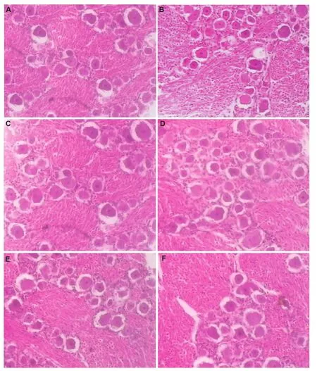
Figure 1 Morphology of trigeminal ganglion cells and axons after percutaneous microballoon compression (hematoxylin-eosin staining, × 100).
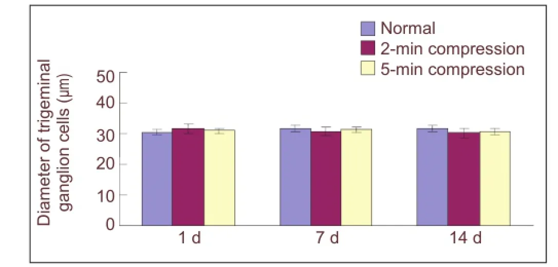
Figure 2 Effect of percutaneous microballoon compression on the diameter of trigeminal ganglion cells.
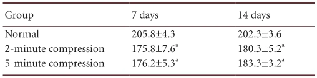
Table 1 Vascular endothelial growth factor immunoreactivity (gray value) in the trigeminal ganglion cell bodies in rabbits at 7 and 14 days after percutaneous microballoon compression
Histomorphological changes in the trigeminal ganglion and in the diameter of ganglion cells after percutaneous microballoon compression
Histological sections of the trigeminal ganglia and roots were prepared from rabbits at 1, 7 and 14 days after percutaneous microballoon compression, and were observed under the light microscope (Figure 1). The trigeminal ganglion cells were analyzed with an image analysis system. Because the myelin sheath contains phospholipids, it is stained negative by hematoxylin-eosin. In comparison, the ganglion cell bodies were round and dyed red, while the nuclei were dyed a deep red. Most perikarya in the ganglion appeared normal and were stained (basophilic). There was no signi fi cant difference in diameter of the trigeminal ganglion cells among the 2-minute compression group, the 5-minute compression group and the normal group (P > 0.05; Figure 2). Thus, the morphology of the trigeminal ganglion cells was not signi ficantly affected by the 2- or 5-minute compression period.
Ultrastructure of the myelin sheath in the trigeminal nerve root after percutaneous microballoon compression
Normal myelin was arranged as homocentric circles, and the layering of the sheath was regular and orderly (Figure 3A). In the 2-minute compression group, the large-diameter myelinated axons were swollen, disrupted and demyelinated 7 days after percutaneous microballoon compression, as revealed under the electron microscope.
Axonal swelling and fragmentation were uniformly present (Figure 3B). At 14 days after compression, fragmentation of myelin and formation of digestion chambers were evident (Figure 3D). In the 5-minute compression group, gross demyelination in the root was detected 7 days after percutaneous microballoon compression. A portion of the myelin had completely disintegrated into fragments, indicating significant demyelination (Figure 3C). At 14 days after compression, some of these fragments had been absorbed, but deformation of the large myelinated fi bers could still be observed (Figure 3E). These changes were not seen in the roots or ganglia in the normal group. These changes are indicative of Wallerian degeneration and retrograde axonal degeneration.
Vascular endothelial growth factor immunoreactivity in trigeminal ganglion cells after percutaneous microballoon compression
There was negative or weak vascular endothelial growth factor immunoreactivity in normal trigeminal ganglion cells. Immunohistochemical staining showed that vascular endothelial growth factor immunoreactivity in the ganglion cell bodies was reduced 7 and 14 days after trigeminal ganglion compression. There was no statistically signi fi cant difference between the 2- and 5-minute compression groups (P > 0.05). In the normal group, vascular endothelial growth factor immunoreactivity in the ganglion cell body was lower than in the compression groups (P < 0.05). These experimental fi ndings indicate that vascular endothelial growth factor immunoreactivity is upregulated following compression (Figure 4; Table 1).
Discussion
Percutaneous microballoon compression was fi rst reported in 1983 by Mullan and Lichtor[20]. Subsequently, it became one of the most widely used alternative therapeutic approaches because of its high (93–99%) initial pain relief[21-25], easy application, and low morbidity[24,26-28]. Unlike glycerol injection and radiofrequency lesioning, which require a cooperative patient[29-30], percutaneous balloon compression is based on timed balloon in fl ation guided with radiographic imaging, and may be performed under general anesthesia.
Because the surgery may be performed under general anesthesia, it is painless to the patient during the entire procedure and is reassuring to the surgeons, irrespective of their familiarity with transforamen ovale long needle techniques. Some researchers[31-34]have proposed that percutaneous microballoon compression is suitable for patients with ophthalmic division involvement, whereas others recommend it as a fi rst-line treatment in patients who are reluctant to have a major surgical operation, such as microvascular decompression. Therefore, percutaneous balloon compression is widely preferred for treating trigeminal neuralgia, especially for elderly patients, because of the high success rate, technical simplicity and relative safety[35-39].
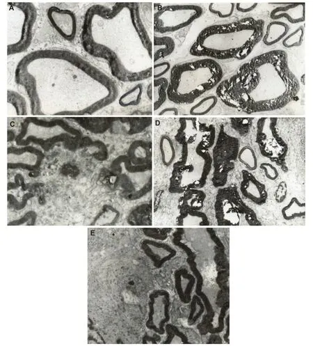
Figure 3 Effect of percutaneous microballoon compression on the ultrastructure of the trigeminal root at 7 and 14 days (d) after percutaneous microballoon compression (transmission electron microscope, × 3,000).
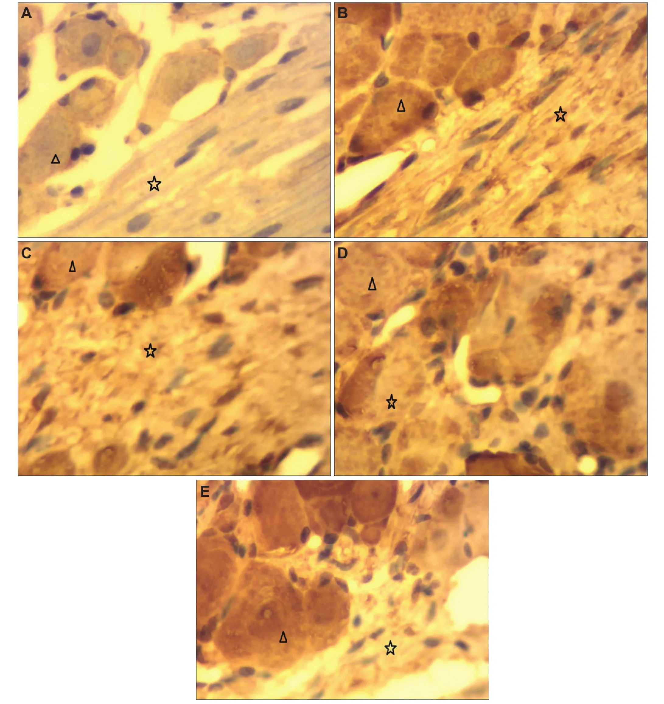
Figure 4 Effect of percutaneous microballoon compression on vascular endothelial growth factor immunoreactivity in trigeminal ganglion cells (immunohistochemical staining, light microscope, × 400).
Percutaneous microballoon compression is generally performed with the guidance of X-ray fluoroscopy[40-45]. This operation requires the use of fluoroscopy to navigate the needle through the foramen ovale and to monitor balloon inflation and deflation. However, the level of radiation exposure for this technique has not been adequately described. Both surgeons and patients are exposed to the radiation. The currently used compression duration ranges from 3 to 6 minutes. Fransen[19]suggested use of lead-coated protective clothing and careful use of the fl uoroscope mandatory.
Scientists[46-48]have modified and reduced the compression time to 2 minutes while maintaining good treatmentoutcome. Our findings indicate that the large myelinated fi bers underwent uniform demyelination under the different periods of compression. Swelling, unraveling, disintegration of myelin and digestion chambers were evident. Gross demyelination in the trigeminal root was detected. These changes were not seen in the roots or ganglia of rabbits in the normal group. These changes represented Wallerian degeneration and retrograde axonal degeneration. There was significant demyelination in the longer-term compression group. For the recurrent patient, an extended duration of compression would be required to obtain a high rate of pain relief and prolong the recurrence interval.
Unfortunately, it remains unclear how percutaneous microballoon compression provides pain relief, although many investigations have been performed[49-50]. This is due, in part, to the complex anatomical structure of the trigeminal nerve. Trigeminal ganglia contain sensory and movement components. The ganglia are formed by a combination of neural crest cells and cells derived from a thickening (placode) of the ectoderm. The trigeminal cell bodies of the first-order trigeminal sensory neurons are located in the semilunar (Gasserian) ganglion of the trigeminal nerve. The fi rst-order neurons for the facial nerve lie in the sensory ganglion for that nerve. The ganglia are, therefore, homologous to the dorsal root ganglia of the spinal nerves. The central processes of the axons of the first-order neurons descend up the brainstem via the spinal trigeminal tract to the spinal trigeminal nucleus, where they synapse with the second-order neurons. The axons of the second-order neurons decussate to the contralateral side and ascend in the ventral trigeminal tract to synapse in the ventral posteromedial nucleus of the thalamus with the third-order neurons. The ventral trigeminal tract or ventral secondary ascending tract of the trigeminal nerve is analogous to the lateral spinothalamic tract. The axons of the third-order neurons pass through the internal capsule to the postcentral gyrus. The entry of the nerve fibers into the spinal tract and nucleus of the trigeminal nerve is highly organized.
It can be described in terms of an imaginary picture of the head being projected onto the nucleus like a slide on a screen. This is called a somatotopic organization and is common in sensory systems. If the nucleus is envisaged as a fi ngerlike object lying parallel to the neuraxis, sensations from successive strips of tissue in the head are fed into successive slices of the nucleus. This is called the onion skin concept. In addition, in a transverse section of that part of the nucleus connected to the trigeminal nerve itself, the three different tiers of cells are selectively associated with the ophthalmic, maxillary and mandibular divisions of the nerve. However, there is little research on the association between the complex anatomical structure and the histopathological changes after percutaneous microballoon compression, especially for different durations of compression. Neurosurgeons from different countries have adopted varying compression periods that range from 3 to 6 minutes[40-45].
To further improve pain relief, many surgical procedures have emerged to analyze the impact of balloon shape, balloon position, balloon volume and duration on the therapeutic effect of percutaneous balloon compression. Our research team modi fi ed the compression time to 2 minutes and obtained satisfactory results[46-48]. Mild to severe facial numbness accompanied by hypoesthesia and transient masticatory weakness was found in most of the patients postoperatively; however, most of these resolved within the fi rst 3–10 months. Corneal anesthesia and keratitis were observed in 1% of patients.
Most technical modifications have been based on the surgeon’s personal experience, with no solid experimental foundation. No histological changes have been reported after acute trigeminal nerve compression. A study of Rydevik and colleagues[51]showed that an external pressure of 2.66 kPa (20 mmHg) reduced epineurial venule blood flow. A pressure of 3.99 kPa (30 mmHg) inhibited both antegrade and retrograde axonal transport, and with 10.64 kPa (80 mmHg) pressure, all endoneurial blood flow ceased. Dyck et al.[52]examined histopathologic changes with acute cuff compression (6.65 kPa [50 mmHg] for 2 minutes) and found an alteration in the shape of the myelin sheath. Szabo and Chidgey[53]and Gelberman et al.[54]demonstrated that pressures within the carpal tunnel were elevated in patients. These authors increased the external pressure within the carpal tunnel for a maximum of 4 hours and studied neurological function. Some neurological functions were lost at 5.32 kPa (40 mmHg), and all functions were blocked at 6.65 kPa (50 mmHg). In volunteers with different blood pressures, the critical extraneural pressure threshold above which function was blocked was 3.99 kPa (30 mmHg) less than the diastolic pressure and 5.98 kPa (45 mmHg) below mean arterial blood pressure. The correlation between systemic blood pressure and neurological function in response to acute compression supports the hypothesis that decreased microvascular blood fl ow plays an important role in nerve compression[55-56].
Conversely, surgeons participating in the randomized trial in Taiwan performed a minimum of 80 D2 LD surgeries before the study began. The results of that study revealed a possible increase in survival rates when extended volumes of LD were performed[35].
What about the histological changes in the trigeminal ganglion after compression injury? In the present study, we performed percutaneous microballoon compression in rabbits with an in fl ation pressure for two different periods of compression (2 and 5 minutes). The results revealed that there was no signi fi cant change in ganglion cell morphology and diameter after percutaneous microballoon compression. However, the larger myelinated fibers were more likely to be injured and demyelinated. These findings are similar to those of Baker and Kerr[28], who performed Shelden’s procedure for ganglion compression through a temporal craniectomy in cats. The demyelination was more severe with prolonged compression. This observation is in accordance with the findings in many patients with a weak zygomatic re fl ex (which is related to the A-alpha fi bers) after percutaneous microballoon compression, which suggests that the compression selectively damages the large myelinated fi bers. This is likely due to the greater sensitivity of large fi bers to mechanical compression. The present study shows that most ganglion cells can withstand compression injury.
Accumulating evidence[57-59]shows that trigeminal root demyelination is the main pathological change in trigeminal neuralgia. However, the mechanism of demyelination remains unclear. Some researchers speculate that it is related to nerve nutrition and metabolism[59]. This would be in ac-cordance with the histopathologic fi ndings of fi brosis, which manifests as a thickening of the external epineurium and perineurium. These changes interfere with blood fl ow in the vessels that pass through the epineurium and perineurium, resulting in ischemia of the nerve fi bers. Numerous experimental studies have demonstrated that nerve injury is related to the degree and duration of compression, with both mechanical and ischemic factors contributing to neurological dysfunction[49-50]. Nukada and Dyck[59]inferred that regional hypoxia causes axonal stasis as a primary event. Demyelination was found in fi bers showing swollen dark and attenuated axons. Their fi ndings suggest that axons are selectively vulnerable to acute ischemia and dependent on its severity; the fi bers either undergo axonal degeneration or transitory structural alterations without axonal degeneration; the latter consisting of axonal changes and secondary demyelination. Kupers et al.[60]showed clear evidence of the demyelination of large myelinated fi bers after compression. Their data indicate that the main histopathological change in the ischemic peripheral nerve after compression is demyelination.
Very little is known of the changes in cytokine levels after percutaneous microballoon compression injury of the trigeminal nerve. A number of researchers[60-62]have shown that vascular endothelial growth factor plays an important role in neural regeneration. Vascular endothelial growth factor is an endothelium-speci fi c growth factor that promotes endothelial cell proliferation, differentiation and survival, mediates endothelium-dependent vasodilatation, induces microvascular hyperpermeability. Hypoxia stimulates the secretion of vascular endothelial growth factor and other angiogenic factors, leading to neovascularization and protection against ischemic injury. Arany et al.[63]showed that vascular endothelial growth factor mRNA activates and controls the progression of angiogenesis. The new vascularization improves blood supply in the ischemic tissue. Ischemic neurons would regain normal function when the oxygen and nutrient supply is restored.
In the present study, there were more vascular endothelial growth factor-positive cells in the compressed ganglion cells, which illustrates that vascular endothelial growth factor expression is upregulated and plays an important role in nerve repair in the early stages of nerve injury. The increased vascular endothelial growth factor expression helps repair the nerve defects. Thus, vascular endothelial growth factor may have a potential as a therapeutic agent for nerve injury repair.
In summary, there were histopathological changes after percutaneous trigeminal ganglion compression in the rabbits, including signi fi cant axon demyelination. High vascular endothelial growth factor expression was also observed. Further experiments are needed to determine if the higher vascular endothelial growth factor levels have a bene fi cial effect.
Materials and Methods
Design
A neuropathological, observational, animal study.
Time and setting
This experiment was performed at the Laboratory of the People’s Hospital of Liaoning Province and the Laboratory Center of Liaoning Medical College, Liaoning, China between April and September 2012.
A total of 36 healthy adult, New Zealand rabbits, aged 12–24 months, of either gender, weighing 2.4–4.3 kg, were provided by the Animal Center of Liaoning Medical College (license No. SCXK (Liao) 2003-0007). The experimental animals were sacri fi ced according to the Guidance Suggestions for the Care and Use of Laboratory Animals, published by the Ministry of Science and Technology of China[64]. All rabbits were housed in an air-conditioned room and were allowed to freely feed.
Methods
Establishment of percutaneous microballoon compression model Percutaneous trigeminal ganglion compression was performed on 30 adult New Zealand White rabbits. After rabbits were anesthetized with ketamine (25 mg/kg intramuscularly), the unilateral foramen ovale and trigeminal ganglion and root were located to determine the optimal percutaneous puncture pathway. An 18-gauage needle (Edwards Lifesciences, Irvine, CA, USA) was positioned at the foramen ovale by fl uoroscopic guidance. When the foramen ovale was identi fi ed on the fl uoroscopic image, a No. 2 Fogarty embolectomy catheter (Edwards Lifesciences) was positioned in Meckel’s cave and advanced 3 to 5 mm. The position of the catheter was confirmed by lateral radiographs (Figure 5). The microballoon was gradually in fl ated with an Omnipaque (Ansheng Corporation, Shanghai, China), with an in fl ation pressure of 1,005 ± 150 mmHg, for 2 or 5 minutes by means of a micrometer (Allwell Medical Corporation, Raleigh, USA) and then removed. The rabbits were intramuscularly injected gentamicin (2.5 mg/kg daily) after percutaneous microballoon compression for 3 days post-surgery to prevent infection.
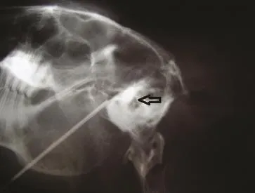
Figure 5 X-ray of the rabbit skull.
Preparation of tissue specimens
At 1, 7 and 14 days after percutaneous microballoon compression, rabbits were deeply anesthetized with 20% ethyl carbamate (1.2 g/kg, intraperitoneal), and transcardially perfused with 500 mL PBS, followed by 500 mL of 4% paraformaldehyde (w/v), and then the trigeminal ganglia were removed. Tissue specimens within 4 mm of the trigeminal ganglion were resected, dehydrated in a graded alcohol series, and paraffin-embedded. The tissue was cut using a Paraffin microtome (Leica, Solms, Germany) into 4-μm-thick sections for hematoxylin-eosin and immunohistochemical staining.
Hematoxylin-eosin staining
The tissue sections were stained in Harris Hematoxylin for 8 minutes. Excess stain was removed by rinsing in running tap water for 2 minutes. Sections were dipped in 0.3% ammonia water or saturated in lithium carbonate water until specimens were jean blue (3–6 dips). Subsequently, tissue sections were rinsed in tap water for 1 hour and immersed in tap water for 2 minutes, then hydrated in a graded alcohol series for 1 minute each and in water for 2 minutes. Alcoholic Eosin Y staining was performed for 2 minutes. Tissue sections were dehydrated in 100% alcohol, placed in xylene, cover-slipped with neutral gum and marked. The morphology of the compressed and normal ganglia and roots was observed under light microscopy (Olympus, Osaka, Japan).
Diameter of trigeminal ganglion cells measured using image analysis
Cell imaging software (Daheng Corporation, Beijing, China) was used to quantify cell diameter. The trigeminal ganglion specimen sections were photographed using an optical microscope (Olympus) at 400 × magnification. The normal ganglion cell structure was clearly visible, and the cell membranes had an even thickness and distinct margins. The total area of the trigeminal ganglion cells was set as a measurement reference. Three sections for each specimen were randomly selected from every group for observation. Four visual fi elds for each section were randomly selected and a sufficient number (≥ 5) of trigeminal cells were measured to reduce error. The dot number of the trigeminal ganglion cell area under a 10 × objective was set as A. According to the systematic setting, 639 dots amount to 400 μm. The acquired area dot A was converted to equivalent diameter dot B. Then, the actual trigeminal ganglion cell diameter was B × 0.626. The mean diameter of every time point after percutaneous microballoon compression was acquired and analyzed statistically. The steps were as follows: (1) Image capture: Sections were placed under the microscope and observed under a 10 × objective lens. Four sections were selected. The image analysis system automatically recorded and displayed three images on the monitor; (2) selection of measurement parameters: The total area of the trigeminal ganglion cells was set as the measurement reference; (3) image enhancement: The obscured and distorted regions were stroked out to enhance the quality of the image by adjusting the contrast and shadow, and by smoothening and sharpening; (4) image segmentation: The gray value was selected based on the image characteristics. Five axon images were selected randomly and converted to binary images; (5) image binary management: The binary image was adjusted to remove objects of no interest; (6) image recognition and analysis process: All images were processed and analyzed according to the instructions provided with the image analysis system, which converted the data to ganglion cell diameter and performed the analysis.
Ultrastructure of the trigeminal ganglion under the electron microscope
The trigeminal ganglion tissues were dissected out quickly and divided into several small pieces (< 1 mm × 1 mm × 1 mm) at low temperature (0–4°C). All the tissues were placed in 2.5% glutaraldehyde fi xing solution for more than 2 hours, rinsed with PBS, fi xed in 1% osmium tetroxide solution for 1.5 hours, pH 7.3–7.4, and rinsed with PBS for 20 minutes. After washing, the tissue was dehydrated with 70% acetone for 15 minutes, 80% acetone for 15 minutes, 90% acetone for 15 minutes, and 100% acetone for 10 minutes twice. Tissues were then placed in a dilute solution of plastic embedding medium epoxy resin mixture, and then polymerized in an oven at 45°C (12 hours), and thereafter at 60°C (36 hours). When the plastic was hard, the block was trimmed for sectioning on the ultramicrotome. Specimens were cut into 50–70-nm thick sections with a glass knife using an LKBNOVE ultramicrotome (Leica). The sections were fl oated off the edge of the knife onto the surface of a water trough, and picked off the surface with a copper grid of 3 mm diameter covered with support Formvar fi lm (10 nm thickness). After the sections were dried, they were stained with heavy metal solutions. Changes in ultrastructure were observed using a JEM-1200EX transmission electron microscope (JEOL, Tokyo, Japan).
Vascular endothelial growth factor immunoreactivity in trigeminal ganglion cells detected by immunohistochemical staining
The tissue specimens were dewaxed and hydrated prior to microwave antigen retrieval in citrate buffer solution, followed by incubation in 3% H2O2to block endogenous peroxidase. After rinsing three times for 5 minutes each, tissue specimens were incubated with normal goat serum for 15 minutes at room temperature, with primary mouse anti-vascular endothelial growth factor polyclone antibody (1:100; Boaosen Corporation, Beijing, China) at 4°C for 24 hours, and with biotinylated goat anti-mouse IgG (1:200; Solarbio Corporation, Beijing, China) for 10 minutes at 37°C. Specimens were then incubated in 3,3′-diaminobenzidine (Beyotime Corporation, Beijing, China) in PBS for 1–2 minutes to reveal labeling. The stained sections were mounted on slides, dehydrated in a graded ethanol series, cleared with xylene, and cover-slipped with neutral gum. Using the Paxinos atlas[65]as a guide, fi ve visual fi elds for each speci fi c region were randomly selected from each of the four sections. The trigeminal ganglion cell bodies and axons labeled brown were positive. Cell imaging software (Daheng Corporation) was used to quantify vascular endothelial growth factor im-munoreactivity, and trigeminal cells and axons were photographed using an optical microscope at 400 × magni fi cation. The ganglion cell bodies and axons in each section were photographed. HPIAS-1000 high definition color image analysis system (Image Engineering Corporation, Wuhan, Hubei Province, China) was used to assay the gray value of each section.
Statistical analysis
All data were expressed as mean ± SD. One-way analysis of variance followed by least signi fi cant difference test was performed using SPSS 10.0 software (SPSS, Chicago, IL, USA). P < 0.05 was considered statistically signi fi cant.
Author contributions:Liu YH and Yi FX checked the manuscript, were in charge of the funds, and provided substantial technical guidance and support. Li FY conducted the experiments and wrote the manuscript. Liu YH and Ma Y revised the paper. Han S, Xu XM, Yi FX and Li FY were responsible for data statistical analyses. All authors approved the final version of the manuscript.
Con fl icts of interest:None declared.
Peer review:Extracellular recording was used to examine response properties in auditory neurons of the inferior colliculus. The results help to explain the difficulty in sound localization and speech perception that patients with hearing aids exhibit in complex sound environments.
[1] Merskey H, Bogduk N. Classi fi cation of Chronic Pain. Descriptors of Chronic Pain Syndromes and De fi nitions of Pain Terms. 2nded. Seattle: IASP Press. 1994.
[2] Sweet WH. Oprative Neurosurgery Techniques: Indications, Methods, and Results. 3rded. Philadelphia: WB Saunders. 1995.
[3] Zakrzewska JM, McMillan R. Trigeminal neuralgia: the diagnosis and management of this excruciating and poorly understood facial pain. Postgrad Med J. 2011;87(1028):410-416.
[4] Headache Classi fi cation Subcommittee of the International Headache Society. The International Classi fi cation of Headache Disorders: 2ndedition. Cephalalgia. 2004;24 Suppl 1:9-160.
[5] Dieleman JP, Kerklaan J, Huygen FJ, et al. Incidence rates and treatment of neuropathic pain conditions in the general population. Pain. 2008;137(3):681-688.
[6] Chen JF, Tu PH, Lee ST. Repeated percutaneous balloon compression for recurrent trigeminal neuralgia: a long-term study. World Neurosurg. 2012;77(2):352-356.
[7] Chen JF, Tu PH, Lee ST. Long-term follow-up of patients treated with percutaneous balloon compression for trigeminal neuralgia in Taiwan. World Neurosurg. 2011;76(6):586-591.
[8] Campos WK, Linhares MN. A prospective study of 39 patients with trigeminal neuralgia treated with percutaneous balloon compression. Arq Neuropsiquiatr. 2011;69(2A):221-226.
[9] Omeis I, Smith D, Kim S, et al. Percutaneous balloon compression for the treatment of recurrent trigeminal neuralgia: long-term outcome in 29 patients. Stereotact Funct Neurosurg. 2008;86(4):259-265.
[10] Skirving JD, Dan NG. A 20-year review of percutaneous balloon compression of the trigeminal ganglion. J Neurosurg. 2001;94(6):913-917.
[11] Brown JA, Pilitsis JG. Percutaneous balloon compression for the treatment of trigeminal neuralgia: results in 56 patients based on balloon compression pressure monitoring. Neurosurg Focus. 2005;18(5):E10.
[12] Jannetta PJ. Treatment of trigeminal neuralgia for micro-operative decompression. In: Youmans JR. Neurological Surgery. Philadelphia: Saunders. 1982.
[13] Kerr FW. Pathology of trigeminal neuralgia: light and electron microscopic observations. J Neurosurg. 1967;26(1):Suppl:151-156.
[14] Natarajan M. Percutaneous trigeminal ganglion balloon compression: experience in 40 patients. Neurol India. 2000;48(4):330-332.
[15] Li YF, Ma Y, Huang HT, et al. Percutaneous microcompression for bilateral trigeminal neuralgia treatment. Jieru Fangshe Xue Zazhi. 2010;19(6):502-504.
[16] Brown JA, McDaniel MD, Weaver MT. Percutaneous trigeminal nerve compression for treatment of trigeminal neuralgia: results in 50 patients. Neurosurgery. 1993;32(4):570-573.
[17] Pickett GE, Bisnaire D, Ferguson GG. Percutaneous retrogasserian glycerol rhizotomy in the treatment of tic douloureux associated with multiple sclerosis. Neurosurgery. 2005;56(3):537-545.
[18] Abdennebi B, Mahfouf L, Nedjahi T. Long-term results of percutaneous compression of the gasserian ganglion in trigeminal neuralgia (series of 200 patients). Stereotact Funct Neurosurg. 1997;68(1-4 Pt 1):190-195.
[19] Fransen P. Fluoroscopic exposure during percutaneous balloon compression of the Gasserian ganglion. J Neurointerv Surg. in press.
[20] Mullan S, Lichtor T. Percutaneous microcompression of the trigeminal ganglion for trigeminal neuralgia. J Neurosurg. 1983;59(6):1007-1012.
[21] Revuelta R, Nathal E, Balderrama J, et al. External carotid artery fi stula due to microcompression of the gasserian ganglion for relief of trigeminal neuralgia. Case report. J Neurosurg. 1993;78(3):499-500.
[22] De Siqueira SR, da Nobrega JC, de Siqueira JT, et al. Frequency of postoperative complications after balloon compression for idiopathic trigeminal neuralgia: prospective study. Oral Surg Oral Med Oral Pathol Oral Radiol Endod. 2006;102(5):e39-45.
[23] Urculo E, Alfaro R, Arrazola M, et al. Trochlear nerve palsy after repeated percutaneous balloon compression for recurrent trigeminal neuralgia: case report and pathogenic considerations. Neurosurgery. 2004;54(2):505-509.
[24] Langford P, Holt ME, Danks RA. Cavernous sinus fi stula following percutaneous balloon compression of the trigeminal ganglion. Case report. J Neurosurg. 2005;103(1):176-178.
[25] Jellish WS, Benedict W, Owen K, et al. Perioperative and long-term operative outcomes after surgery for trigeminal neuralgia: microvascular decompression vs percutaneous balloon ablation. Head Face Med. 2008;4:11.
[26] Lopez BC, Hamlyn PJ, Zakrzewska JM. Systematic review of ablative neurosurgical techniques for the treatment of trigeminal neuralgia. Neurosurgery. 2004;54(4):973-983.
[27] Sweet WH, Polleti CE. Complications of percutaneous rhizotomy and microvascular decompression operations for facial pain. In: Schmidek H, Sweet W, eds. Current Techniques in Operative Neurosurgery. Orlando: Grune & Stratton. 1988.
[28] Baker GS, Kerr FW. Structural changes in the trigeminal system following compression procedures. J Neurosurg. 1963;20:181-184. [29] Kanpolat Y, Ugur HC. Systematic review of ablative neurosurgical techniques for the treatment of trigeminal neuralgia. Neurosurgery. 2005;57(3):E601.
[30] Hannerz J, Linderoth B. Neurosurgical treatment of short-lasting, unilateral, neuralgiform hemicrania with conjunctival injection and tearing. Br J Neurosurg. 2002;16(1):55-58.
[31] Sweet WH. The treatment of trigeminal neuralgia (tic douloureux). N Engl J Med. 1986;315(3):174-177.
[32] Broggi G, Ferroli P, Franzini A. Treatment strategy for trigeminal neuralgia: a thirty years experience. Neurol Sci. 2008;29 Suppl 1: S79-82.
[33] Liu HB, Ma Y, Zou JJ, et al. Percutaneous microballoon compression for trigeminal neuralgia. Chin Med J (Engl). 2007;120(3):228-230.
[34] Lee ST, Chen JF. Percutaneous trigeminal ganglion balloon compression for treatment of trigeminal neuralgia, part II: results related to compression duration. Surg Neurol. 2003;60(2):149-154.
[35] Bergenheim AT, Linderoth B. Diplopia after balloon compression of retrogasserian ganglion rootlets for trigeminal neuralgia: technical case report. Neurosurgery. 2008;62(2):E533-534.
[36] Lobato RD, Rivas JJ, Sarabia R, et al. Percutaneous microcompression of the gasserian ganglion for trigeminal neuralgia. J Neurosurg. 1990;72(4):546-553.
[37] Lee ST, Chen JF. Percutaneous trigeminal ganglion balloon compression for treatment of trigeminal neuralgia-part I: pressure recordings. Surg Neurol. 2003;59(1):63-67.
[38] Brown JA. Percutaneous trigeminal nerve compression. In: Batjer HH, Loftus CM, eds. Textbook of Neurological Surgery: Principles and Practice. Philadelphia: Lippincott Williams & Wilkins. 2002.
[39] Wilkinson HA. Percutaneous trigeminal ganglion balloon compression for treatment of trigeminal neuralgia--part I: pressure recordings. Surg Neurol. 2003;60(5):470.
[40] Trojnik T, migoc T. Percutaneous trigeminal ganglion balloon compression rhizotomy: experience in 27 patients. Scienti fi cWorldJournal. 2012;2012:328936.
[41] Chen JF, Lee ST. Comparison of percutaneous trigeminal ganglion compression and microvascular decompression for the management of trigeminal neuralgia. Clin Neurol Neurosurg. 2003;105(3):203-208.
[42] Correa CF, Teixeira MJ. Balloon compression of the Gasserian ganglion for the treatment of trigeminal neuralgia. Stereotact Funct Neurosurg. 1998;71(2):83-89.
[43] Goerss SJ, Atkinson JL, Kallmes DF. Variable size percutaneous balloon compression of the gasserian ganglion for trigeminal neuralgia. Surg Neurol. 2009;71(3):388-391.
[44] Park SS, Lee MK, Kim JW, et al. Percutaneous balloon compression of trigeminal ganglion for the treatment of idiopathic trigeminal neuralgia: experience in 50 patients. J Korean Neurosurg Soc. 2008;43(4):186-189.
[45] Abdennebi B, Amzar Y. Treatment of essential trigeminal neuralgia by Gasserian compression using balloon (50 cases). Neurochirurgie. 1991;37(2):115-118.
[46] Li FY, Ma Y, Zou JJ, et al. Related pain free after percutaneous microcompression treatment for trigeminal neuralgia. Zhongguo Wei Qinxi Shenjing Waike Zazhi. 2010;15(3):105-106.
[47] Wang B, Ma Y, Zou JJ, et al. Clinical experience of percutaneous microcompression treatment for trigeminal neuralgia. Zhonghua Shenjing Waike Zazhi. 2008;24(5):330.
[48] Li YF, Ma Y, Zou JJ, et al. Percutaneous microcompression for trigeminal neuralgia treatment. Zhongguo Wei Qinxi Shenjing Waike Zazhi. 2008;13(11):514-515.
[49] Hilton DA, Love S, Gradidge T, et al. Pathological fi ndings associated with trigeminal neuralgia caused by vascular compression. Neurosurgery. 1994;35(2):299-303.
[50] Devor M, Govrin-Lippmann R, Rappaport ZH. Mechanism of trigeminal neuralgia: an ultrastructural analysis of trigeminal root specimens obtained during microvascular decompression surgery. J Neurosurg. 2002;96(3):532-543.
[51] Rydevik B, Lundborg G, Bagge U. Effects of graded compression on intraneural blood blow. An in vivo study on rabbit tibial nerve. J Hand Surg Am. 1981;6(1):3-12.
[52] Dyck PJ, Lais AC, Giannini C, et al. Structural alterations of nerve during cuff compression. Proc Natl Acad Sci U S A. 1990;87(24):9828-9832.
[53] Szabo RM, Chidgey LK. Stress carpal tunnel pressures in patients with carpal tunnel syndrome and normal patients. J Hand Surg Am. 1989;14(4):624-627.
[54] Gelberman RH, Szabo RM, Williamson RV, et al. Sensibility testing in peripheral-nerve compression syndromes. An experimental study in humans. J Bone Joint Surg Am. 1983;65(5):632-638.
[55] Jeon HJ, Han SR, Park MK, et al. A novel trigeminal neuropathic pain model: compression of the trigeminal nerve root produces prolonged nociception in rats. Prog Neuropsychopharmacol Biol Psychiatry. 2012;38(2):149-158.
[56] Mackinnon SE. Pathophysiology of nerve compression. Hand Clin. 2002;18(2):231-241.
[57] Kerr FW. Evidence for a peripheral etiology of trigeminal neuralgia. 1967. J Neurosurg. 2007;107(1):225-231.
[58] Zhou JH, Jiang XZ, Sun WG, et al. The correlation between Trigeminal nerve demyelination and pathological change of intraneural microcirculation. Xiandai Kouqiang Yixue Zazhi. 2004;18 (6):483-485.
[59] Nukada H, Dyck PJ. Acute ischemia causes axonal stasis, swelling, attenuation, and secondary demyelination. Ann Neurol. 1987;22(3):311-318.
[60] Kupers R, Yu W, Persson JK, et al. Photochemically-induced ischemia of the rat sciatic nerve produces a dose-dependent and highly reproducible mechanical, heat and cold allodynia, and signs of spontaneous pain. Pain. 1998;76(1-2):45-59.
[61] Wang LX, Yin RX, Sun JB. Effect of Tongxinluo on nestin and vascular endothehal growth factor mRNA expression in rat brain tissue after cerebral ischemia-reperfusion injury. Nan Fang Yi Ke Da Xue Xue Bao. 2008;28(12):2131-2135.
[62] Hall GC, Carroll D, Parry D, et al. Epidemiology and treatment of neuropathic pain: the UK primary care perspective. Pain. 2006;122(1-2):156-162.
[63] Arany Z, Foo S Y, Ma Y, et al . HIF-independent regulation of VEGF and angiogenesis by the transcriptional coactivator PGC-1alpha. Nature. 2008;451(7181):1008-1012 .
[64] The Ministry of Science and Technology of the People’s Republic of China. Guidance Suggestions for the Care and Use of Laboratory Animals. 2006-09-30.
[65] Urban I, Richard P. A Stereotaxic Atlas of the New Zealand Rabbit’s Brain. Spring fi eld, IL: Charles C Thomas Publisher Ltd. 1972.
Copyedited by Patel B, Stow A, Norman C, Zou JJ, Wang LM, Yang Y, Li CH, Song LP; Liu WJ, Zhao M
10.4103/1673-5374.125347
Yunhui Liu, Ph.D., Professor, Department of Neurosurgery, Shengjing Hospital, China Medical University, Shenyang 110004, Liaoning Province, China, 13998892068@126.com.
http://www.nrronline.org/
Accepted: 2013-10-10
- 中國神經(jīng)再生研究(英文版)的其它文章
- Acupuncture/electroacupuncture enhances antidepressant effect of Seroxat: the Symptom Checklist-90 scores
- Effects of Wen Dan Tang on insomnia-related anxiety and levels of the brain-gut peptide Ghrelin
- Role of the nucleus tractus solitarii in the protection of pre-moxibustion on gastric mucosal lesions
- Mild hypothermia for treatment of diffuse axonal injury: a quantitative analysis of diffusion tensor imaging
- The optimal distance between two electrode tips during recording of compound nerve action potentials in the rat median nerve
- Acupuncture at Baihui and Dazhui reduces brain cell apoptosis in heroin readdicts