
Auricular embryonal rhabdomyosarcoma
2022-06-28 12:22:34JavierArredondoMonteronicaBronteAnautSaraHernndezMartJuanCarlospezGutirrez
World Journal of Pediatrics 2022年5期
Javier Arredondo Montero ·Mónica Bronte Anaut·Sara Hernández-Martín·Juan Carlos López-Gutiérrez
A 13-month-old girl presented with a red-violaceous tumor of 13 mm in diameter on the inner side of the root of the right helix,with increased temperature and cystic consistency (Fig.1).No pulse was palpable in the lesion.
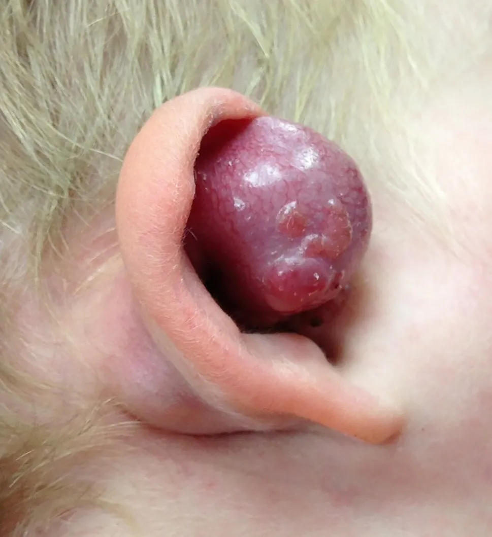
Fig.1 Clinical photograph of the patient.A red-violaceous tumor of 13 mm in diameter is identified in the internal face of the root of the helix.Retroauricular tumor is diagnosed
An ultrasonography was performed,showing a 1.6 cm rounded lesion with well-defined margins,hypoechogenic and homogeneous,compatible with hemangioma.Empirical treatment with oral propranolol was started without improvement.During the following weeks,progressive growth of the lesion was observed and was accompanied by the appearance of subcutaneous nodular lesions at the preauricular level.At the same time,the patient presented spontaneous self-limited bleeding from the lesion that required admission for dressings and monitoring.The episode resolved without incident.
An MRI was requested,showing a polylobulated mass at the right facial level involving auricular pavilion,retroauricular space,parotid,retroparotid and lateral cervical regions (Fig.2).A biopsy was performed,with the diagnosis of embryonal rhabdomyosarcoma.An extensive study was performed with no signs of distant malignancy.Surgical resection was indicated.
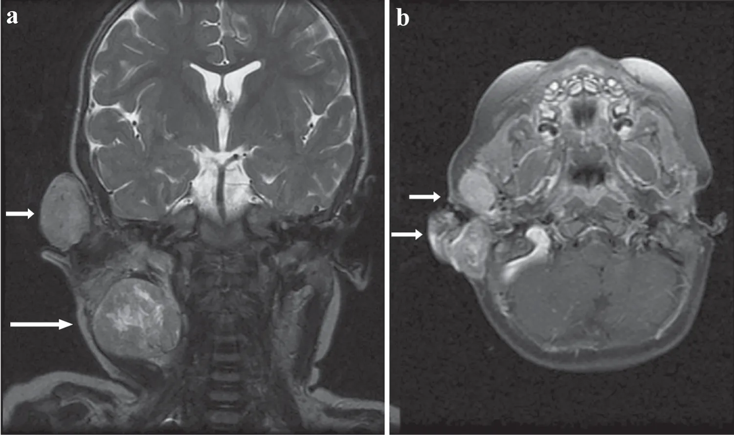
Fig.2 Faciocervical MRI.a Coronal section; b Axial section.A polylobulated mass at the right facial level involving auricular pavilion,retroauricular space,parotid,retroparotid and lateral cervical regions is identified (white arrows)
The anatomopathological study of the specimen showed that it was a stage IIC embryonal rhabdomyosarcoma,with an affected resection margin.Three infiltrated lymph nodes also were identified at the jugulodigastric level.The patient received adjuvant treatment with chemotherapy and radiotherapy.The patient is currently 8 years old,is under evolutionary follow-up and is asymptomatic without oncologic recurrence.
Rhabdomyosarcoma is an aggressive neoplasm in the pediatric population.It is classified mainly into two variants,alveolar and embryonal [1].Auricular presentation is extremely rare,being only isolated pediatric reports in the scientific literature,most of them being of embryonal subtype [2–7].Patients usually present with a progressively enlarging auricular or retroauricular tumor that is associated with local discomfort,bleeding or discharge through the ear canal,as well as hearing loss.Good prognostic factors are considered to be age less than 10 years,embryonal histological subtype,complete primary resection,absence of locoregional adenopathic involvement and tumor size less than 5 cm [4].Despite its vascular appearance,which can be confused with a vascular anomaly,such as hemangioma or lymphangioma [7],the presence of a fast-growing tumor at the auricular level should lead us to consider the diagnosis of rhabdomyosarcoma.Ultrasound generally shows non-specific findings,so if there is a diagnostic suspicion,other imaging modalities,such as MRI,should be used.Individualized therapeutic guidance is required for each patient.
AcknowledgementsTo the patient's parents,for their willingness and collaboration in publishing this case report.
Author contributionsJAM,MBA:conceptualization and study design;literature search and selection,data curation and extraction,formal analysis;investigation;methodology;project administration;resources;validation;visualization;writing—original draft;writing—review and editing.SHM,JCLG:formal analysis;investigation;writing—original draft;writing—review and editing.All authors listed have made a substantial,direct and intellectual contribution to the work,and approved it for publication.
FundingThere is no external funding to declare.
Data availabilityNo data for this clinical image.
Declarations
Ethical approvalThis study was exempt from formal evaluation by the Clinical Research Ethics Committee of our center,because it was an isolated retrospective clinical report of a descriptive nature.
Conflict of interestNo financial benefits have been received or will be received from any party related directly or indirectly to the subject of this article.C
onsent for publicationPrior to the submission of this article,verbal and written informed consent was obtained from the legal guardians of the patient whose clinical photographs are included in this publication.The patients' medical records were accessed in accordance with the specific hospital regulations applicable to this type of case.
 World Journal of Pediatrics2022年5期
- World Journal of Pediatrics的其它文章
- Instructions for Authors
- Editors
- Unrecognized congenital heart disease in rural school-age children:getting to the root of the problem
- Insufficient application of estimation equations of glomerular filtration rate:a survey of 1009 Chinese pediatricians
- Schoolchildren from disadvantaged backgrounds present a loss of lean tissue mass and significant increase of body fat mass during the COVID-19 lockdown in Germany:results from the MEDdirect study
- Unusual cause of recurrent vomiting with failure to thrive in an infant